Falsifiable
designating or of a statement, theory, etc. that is so formulated as to permit empirical testing and, therefore, can be shown to be false
https://www.collinsdictionary.com/us/dictionary/english/falsifiable
Beyond adherence to the scientific method, a key factor in determining whether or not the evidence gathered during research is indeed scientific rather than pseudoscientific is a simple concept known as falsifiability. This idea was introduced by scientific philosopher Karl Popper in 1935 in his book The Logic of Scientific Discovery. Essentially, what falsifiability means is that, in order for a hypothesis or theory to be scientific, it must have the abilty to be disproven. Someone should be able to conceivably design an experiment that could prove the hypothesis or theory wrong. If a hypothesis or theory is capable of being proven wrong and yet it is supported by experimental evidence of its truth, then it can be considered as a scientific hypothesis or theory.
Popper explained his reasoning for this criterion in his 1963 book Conjectures and Refutations. Falsification was an attempt to draw a line between science and pseudoscience. He was bothered by the ways in which observations could be easily fit to confirm whatever theory was believed by the eye of the beholder. In this way, it is easy to create a confirmation bias where one ignores contradictory information in order to claim that the vast majority of the observations fit within their own theoretical paradigm. Thus, Popper laid out some rules that could discern real scientific confirmations over those that are pseudoscientific. Confirmations should only be accepted if they are able to be refuted. If the hypothesis or theory stands tall in face of the experiments attempting to disprove it, this gives the researcher stronger conviction that their hypothesis and/or theory is correct. Popper wanted to ensure that those who believed in a certain hypothesis or theory could not reinterpret it in a way that it could escape refutation once it was proven to be false:
“The most characteristic element in this situation seemed to me the incessant stream of confirmations, of observations which "verified" the theories in question; and this point was constantly emphasize by their adherents. A Marxist could not open a newspaper without finding on every page confirming evidence for his interpretation of history; not only in the news, but also in its presentation — which revealed the class bias of the paper — and especially of course what the paper did not say. The Freudian analysts emphasized that their theories were constantly verified by their "clinical observations." As for Adler, I was much impressed by a personal experience. Once, in 1919, I reported to him a case which to me did not seem particularly Adlerian, but which he found no difficulty in analyzing in terms of his theory of inferiority feelings, Although he had not even seen the child. Slightly shocked, I asked him how he could be so sure. "Because of my thousandfold experience," he replied; whereupon I could not help saying: "And with this new case, I suppose, your experience has become thousand-and-one-fold."
What I had in mind was that his previous observations may not have been much sounder than this new one; that each in its turn had been interpreted in the light of "previous experience," and at the same time counted as additional confirmation. What, I asked myself, did it confirm? No more than that a case could be interpreted in the light of a theory. But this meant very little, I reflected, since every conceivable case could be interpreted in the light Adler's theory, or equally of Freud's. I may illustrate this by two very different examples of human behavior: that of a man who pushes a child into the water with the intention of drowning it; and that of a man who sacrifices his life in an attempt to save the child. Each of these two cases can be explained with equal ease in Freudian and Adlerian terms. According to Freud the first man suffered from repression (say, of some component of his Oedipus complex), while the second man had achieved sublimation. According to Adler the first man suffered from feelings of inferiority (producing perhaps the need to prove to himself that he dared to commit some crime), and so did the second man (whose need was to prove to himself that he dared to rescue the child). I could not think of any human behavior which could not be interpreted in terms of either theory. It was precisely this fact—that they always fitted, that they were always confirmed—which in the eyes of their admirers constituted the strongest argument in favor of these theories. It began to dawn on me that this apparent strength was in fact their weakness.”
“These considerations led me in the winter of 1919-20 to conclusions which I may now reformulate as follows.
1. It is easy to obtain confirmations, or verifications, for nearly every theory — if we look for confirmations.
2. Confirmations should count only if they are the result of risky predictions; that is to say, if, unenlightened by the theory in question, we should have expected an event which was incompatible with the theory — an event which would have refuted the theory.
3. Every "good" scientific theory is a prohibition: it forbids certain things to happen. The more a theory forbids, the better it is.
4. A theory which is not refutable by any conceivable event is non-scientific. Irrefutability is not a virtue of a theory (as people often think) but a vice.
5. Every genuine test of a theory is an attempt to falsify it, or to refute it. Testability is falsifiability; but there are degrees of testability: some theories are more testable, more exposed to refutation, than others; they take, as it were, greater risks.
6. Confirming evidence should not count except when it is the result of a genuine test of the theory; and this means that it can be presented as a serious but unsuccessful attempt to falsify the theory. (I now speak in such cases of "corroborating evidence.")
7. Some genuinely testable theories, when found to be false, are still upheld by their admirers — for example by introducing ad hoc some auxiliary assumption, or by reinterpreting the theory ad hoc in such a way that it escapes refutation. Such a procedure is always possible, but it rescues the theory from refutation only at the price of destroying, or at least lowering, its scientific status. (I later described such a rescuing operation as a "conventionalist twist" or a "conventionalist stratagem.")
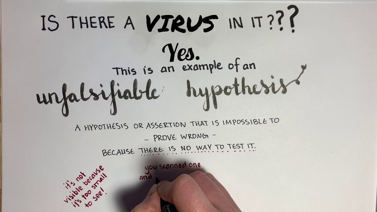
To understand the concept of falsifiability better, let's briefly examine what would be considered a falsifiable versus an unfalsifiable hypothesis. A falsifiable hypothesis could be stated as such:
If I water my plant every day, then it will grow.
This hypothesis can be easily tested to determine if watering the plant every day will, in fact, help it grow. It is falsifiable as it is possible that watering the plant every day will actually cause the plant not to grow as it may actually die from being oversaturated with too much water. If this latter scenario is observed, this would mean that the alternative hypothesis is proven to be false.
On the other hand, an extreme example of what would be an unfalsifiable hypothesis is blaming invisible unicorns for observing a ball falling to the ground as described here:
How we edit science part 1: the scientific method
“An untestable hypothesis would be something like “the ball falls to the ground because mischievous invisible unicorns want it to”. If these unicorns are not detectable by any scientific instrument, then the hypothesis that they’re responsible for gravity is not scientific.
An unfalsifiable hypothesis is one where no amount of testing can prove it wrong. An example might be the psychic who claims the experiment to test their powers of ESP failed because the scientific instruments were interfering with their abilities.”
https://theconversation.com/how-we-edit-science-part-1-the-scientific-method-74521
Of course, unicorns have never been proven to exist and we have no ability to actually observe, study, and detect them. While there may be hundreds or even thousands of indirect confirmatory evidence supporting this unicorn hypothesis, there is no ability to actually determine any truth in regard to the initial hypothesis as there is no ability to test it without unicorns existing. Thus, this is an unfalsifiable hypothesis.
It may already be evident based upon the unicorn analogy that “viruses” actually fit into this same unfalsifiable mold. As these entities have never been observed in nature and directly studied, there is no abilty to know how to detect and actually test for these invisible “pathogens.” The concept of a “virus” was created in order to explain symptoms of disease that could not be pinned on bacteria. However, as with bacteria, pseudoscientific rules were established in the face of contradictory experimental evidence in order to trick the masses into believing that the field was scientific when, in fact, it is anything but. Let's explore some of these pseudoscientific concepts more in depth and see just how unfalsifiable virology, and by extension germ theory, truly is.
Unfalsifiable Concept # 1: Asymptomatic Disease
When Robert Koch was hard at work uncovering his evidence for “pathogenic” bacteria, he established a series of Postulates, or logic-based criteria, that were accepted as true in order to further investigate the potential of disease-causing microbes. The very first of these Postulates required that the microorganism assumed to be the cause of a particular disease should be found in all individuals suffering from a very specific set of symptoms being investigated, and that the microorganism should not be present in those who are free of these symptoms. This very first Postulate set up a falsifiable concept in that one could disprove a specific microbe as being the presumed pathogenic agent if it was found in those who were without illness as well as not being found in those with the illness.
However, after gaining prestige and accolades for his Postulates as well as for his work with tuberculosis, Koch began to realize from further investigation that the microbes he had blamed as the pathogenic agents were, in fact, regularly found in those who were healthy. He uncovered this fact as he attempted to discover a bacterial cause of cholera. In many instances, the comma-shaped bacilli that Koch had championed as the cause of cholera were regularly found in those who are healthy. There were also many cases where the cholera symptoms were present but the bacteria was never found. As he had staked his reputation on discovering the cause of cholera, Koch ended up abandoning the logic of his first Postulate in order to allow for the microbe not to be present in all cases of the disease as well as to be found in those who are healthy:
On the current status of bacteriological cholera diagnosis
“This is not to say, however, that conversely the absence or rather the non-detection of cholera bacteria in a case suspected of having cholera proves the absence of cholera under all circumstances. Just as with other infectious diseases caused by microorganisms, there can also be isolated cases of cholera which, because of their behavior in other respects, must be regarded as indubitable cases of cholera, but in which cases, either because the investigator is insufficiently qualified or because they were examined at an unsuitable point in time are, the cholera bacteria are not found.”
“These mildest cases of cholera, in which cholera bacteria have been found in the solid deposits of apparently healthy people, occur only among groups of people who have been equally exposed to the infection and who show severe cases as well as the mild ones. Nothing of the kind has ever been found in persons who could not possibly have been infected. One must therefore regard these cases as real cases of cholera and cannot use them as evidence against the specific character of the cholera bacteria.”
https://link.springer.com/article/10.1007/BF02284324
Koch's abandonment of his first Postulate set up the first unfalsifiable premise for both germ theory and virolgy. The microbe did not have to be present in all cases of the disease and it was allowed to be present in those who are healthy. As such, there is no way to disprove vibrio cholerae as a pathogenic agent. This unfalsifiable premise was well-known even during Koch's time, as demonstrated by a 1895 letter to the editor from Henry Raymond Rogers, M.D.
Dr. Robert Koch and His Germ Theory of Cholera.
“But this comma-shaped bacillus theory of cholera has proved a failure. These invisible comma-shaped germs are now found to be universal and harmless. They are found in the secretions of the mouth and throat of healthy persons, and in the common diarrheas of summer everywhere—they swarm in the intestines of the healthy and are observed in hardened fecal discharges as well. Dr. Koch today asserts that these bacilli are universally present. He even tells us that: "Water from whatever source frequently, not to say invariably, contains comma-shaped organisms."
Drs. Pettenkofer of Munich and Emmerich of Berlin, physicians of high distinction and experts in this disease, drank each a cubic centimeter of "culture broth" which contained these bacilli, without experiencing a single symptom characteristic of cholera, although the draught in each instance was followed by liquid stools swarming with these germs.
Dr. Koch has kept au courant with the foregoing facts, as well as others quite as significant, and, had he accepted the evidences which thus year after year have been forced upon him, his pernicious cholera germ theory with its most disastrous consequences in misleading mankind would have been unknown today.”
Henry Raymond Rogers, M.D.
https://jamanetwork.com/journals/jama/article-abstract/453342
Rogers and others alerted Koch to the fact that the bacilli he had claimed was the causative agent of cholera were, in fact, regularly found in the water and in healthy people. It had been demonstrated not to be pathogenic via ingestion by well-respected physicians. Even Koch eventually tested the bacilli on himself and was unsuccessful in demonstrating pathogenicity. However, even though he could not satisfy his very first Postulate (as well as the latter 3), Koch allowed for the creation of the unfalsifiable concept of the asymptomatic carrier of disease, establishing both germ theory and virology as a pseudoscience from the very start.
Unfalsifiable Concept # 2: Antibodies & The Immune System
In 1918, Milton Rosenau attempted to find the causative agent of the dreaded Spanish flu that was claimed to be ravaging the world at the time. In order to do so, Rosenau enlisted 100 volunteers with no history of influenza for a study conducted on Gallops Island in Boston. Similar experiments were carried out at Angel Island on the west coast as well. During the initial part of the experiment, the volunteers were subjected to one strain and then several strains of Pfeiffer’s bacillus by spray and swab into their noses, throats and into their eyes. This bacillus was the initial presumed cause and every attempt to transmit it failed. Once the bacterium was ruled out, the volunteers were exposed to other microorganisms obtained from the nose and throats of the influenza victims. When this proved to be unsuccessful, the blood from infected patients was injected into the volunteers which also resulted in a failure to transmit the disease. Finally, 13 volunteers were taken into an influenza ward and exposed to 10 influenza patients each. The volunteers were told to shake hands with each influenza patient and to have conversations as close as possible. The patient was to exhale as hard as he could while the volunteers breathed in. The volunteers were then instructed to permit the sick patient to cough directly into their face. Each volunteer repeated this process with 10 different influenza patients. None of the volunteers on either coast became sick. This experiment led Rosenau to conclude that he had no knowledge about how influenza spread:
“As a matter of fact, we entered the outbreak with a notion that we knew the cause of the disease, and were quite sure we knew how it was transmitted from person to person. Perhaps, if we have learned anything, it is that we are not quite sure what we know about the disease.“
https://zenodo.org/record/1505669/files/article.pdf?download=1
These experiments, during the height of what is considered the most deadly and contagious “virus” of all time, should be enough to convince anyone thinking both critically and logically that the contagion hypothesis was successfully disproven. In every which way conceivablely possible, Rosenau could not transmit the invisible “virus” from the sick to the healthy. Thus, the hypothesis that the fluids of a sick patient contain a “virus” that could transmit disease to a healthy host was falsified.
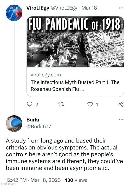
However, this has not stopped germ theory defenders from creating an unfalsifiable excuse in an attempt to disregard these findings. While I have seen ridiculous claims that we can not rely on studies from over 100 years ago as if age somehow invalidates the evidence, one of the more recent excuses was that the immune system was not factored into these studies.
According to Martin, as influenza was unknown, Rosenau did not perform the necessary serological and virological assays. A similar sentiment was presented by someone going by Burki as well, along with the added caveat of the asymptomatic carrier concept thrown in for good measure.
What this line of argument is attempting to do is discredit Rosenau's findings by claiming that the researchers did not check to see if these patients had antibodies to the influenza “virus” and were unknowingly immune to the “virus.” They want people to believe this even though none of the volunteers had a history of “infection.” They argue that perhaps the volunteers were “infected” but were asymptomatic carriers and did not realize it. This line of attack is a barrage of unfalsifiable concepts.
However, even “knowing” today what the influenza “virus” is and how antibodies are said to respond to it has not stopped the confusion in regard to what these readings mean and at what level they supposedly offer “protection.” In a 2013 study looking at vaccine-induced antibody responses, it was made clear that determining a correlate of protection for influenza mostly amounted to guesswork:
Complex Correlates of Protection After Vaccination
Influenza
“The multiple correlates of protection that have been proposed for influenza vaccines provide a perfect example of the complexity of the subject [14]. Clearly, serum antibody is an important mCoP, as measured either by hemagglutination inhibition (HAI) or microneutralization and plays a role in clearance as well as prevention of acquisition [15, 16]. Although an HAI titer of 1:40 has been taken by regulatory authorities as the level that will protect most people, there is disagreement as to whether that number is correct, and in any case protection seems to be a continuous function, with higher titers giving higher levels of protection [17]. In a study of a cell-culture produced vaccine, a titer of 1:15 was claimed to be adequate [18], although to this reader it appeared that 1:30 was a safer bet. On the other hand, in children it has been claimed that a titer of 1:110 is necessary [19]. In another study, some doubt was expressed about the value of serum antibody, but the data appeared to support the protective value of titers >1:32 or >1:64 [20]. Thus, although no serum antibody titer is completely protective in itself, nevertheless the 1:40 titer appears to be a reasonable statistical correlate for an efficacy of 50%–70% against clinical symptoms of infection.”
https://academic.oup.com/cid/article/56/10/1458/402211
Keep in mind that these studies are performed under highly controlled conditions, yet the results meant to determine what antibody measurement equalled “protection” ranged from 1:15 to 1:110. It was settled that 1:40 was a “reasonable statistical correlate” while the researchers admitted that no serum antibody titre is completely protective in and of itself. Thus, these antibody measurements offer nothing that can dispute the Rosenau findings.
However, even if Rosenau had access to the serological assays and tests that we have today, what would this ultimately have told him? How would it have changed the experiment in any way? Highlights taken from a July 2021 study show exactly how little we actually “know” about antibody results for influenza. For most of its history, the antibody results for influenza was derived from hemagglutinin (HA) protein. However, the authors noted that growing evidence pointed to limitations with this approach. The issue of antibody non-responsiveness after both infection and vaccination was an issue that was deemed necessary to explore. In other words, there are cases where people are “infected'“ with and/or vaccinated against the influenza “virus” who failed to produce any measurable antibody response. This creates the unfalsifiable concept where antibodies are allowed to be both present and absent in cases of previous “infection” and vaccination. Instead of realizing that the antibody hypothesis has been falsified from these findings, the researchers attempted to find ways to explain away why they are unable to observe a response in individuals where one should have occurred.
The authors noted that “non-neutralizing” antibodies are regularly detected in the population, and yet, it is unknown if these relate to protection as this finding is vastly understudied. It is admitted as well that seroconversion was typically based on HAI-antibody titer increases as this was the cornerstone of influenza diagnosis before the advent of molecular techniques. However, PCR and other methods have shown that influenza “infections” do not always result in seroconversions, i.e. signs that the immune system is reacting to the presence of the “virus” in the body. The correlation between the initial infection dose, disease severity, and antibody development is admitted to be a challenge to address in any “naturally-acquired infection” due to difficulties in determining the time of “infection.” Thus, the antibody response in humans is not well-established and can only be inferred from animal studies:
Antibody Responsiveness to Influenza: What Drives It?
“The induction of a specific antibody response has long been accepted as a serological hallmark of recent infection or antigen exposure. Much of our understanding of the influenza antibody response has been derived from studying antibodies that target the hemagglutinin (HA) protein. However, growing evidence points to limitations associated with this approach. In this review, we aim to highlight the issue of antibody non-responsiveness after influenza virus infection and vaccination.”
“In summary, non-neutralizing antibodies can be detected in the population, seemingly accumulate with age, and may contribute to protection against newly emerging influenza viruses, although the degree of in vivo protection in humans has not yet been formally established.”
“In the case of influenza, seroconversion would typically be based on HAI-antibody titer increases, which was the cornerstone of influenza diagnosis before the advent of molecular techniques. However, the use of both PCR-diagnosis and serology in large seroepidemiological and human challenge studies has indicated that influenza virus infections do not always result in seroconversions (Table 2).”
“Overall, the correlation between the initial infection dose, disease severity, and antibody development is a challenge to address in any naturally-acquired infection studies due to difficulties in determining the time of infection. Although the time and infection doses are predetermined in LAIV and human challenge studies, for ethical considerations, the viruses used are usually attenuated, or inoculated at doses that do not induce significant symptoms. As such, the relationship between the initial infection dose, symptom severity, and the subsequent downstream antibody response in humans is not well-established and can only be inferred from animal studies.”
“Studying the immunogenicity of the influenza virus is complicated due to the large diversity of antigenic variants present in nature and our constant exposure to it, either through natural infection or vaccination. We have attempted to provide an up-to-date, although by no means comprehensive overview of factors that may influence influenza antibody responses and our ability to measure it (summarized in Figure 1).”
https://www.ncbi.nlm.nih.gov/pmc/articles/PMC8310379/
Ultimately, what these highlights show is that, even with the assays and technology available today and with the backing of decades of research, had Rosenau measured antibody levels in his volunteers, it would have been meaningless. This is especially true as there are cases of the previously “infected” and vaccinated eliciting zero antibody response. Thus, taking into account antibody measurements would have been a pointless endeavor that would make no difference in the outcome of the study as, according to the pseudoscientific narrative, there are people who were “infected” with antibody responses and those who were “infected” without one. There is no way to tell, based on antibody measurements, who has been “infected” and has “immunity,” and who does not. All this does is give researchers a fraudulent escape clause for why they get results that falsify their hypothesis.
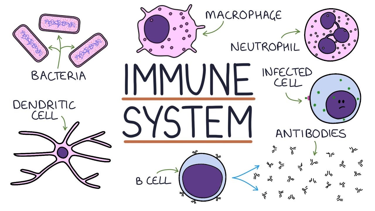
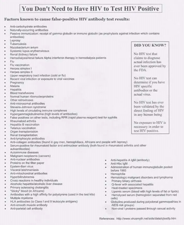
Beyond the issues with correlates of protection and the inability to detect antibodies in those who are “infected” and vaccinated, anyone looking into antibodies seriously will come to the realization that there are other major issues with these entities as well. For instance, if antibodies for HIV are detected, this does not mean that you have successfully fought off the “virus” and are now “protected” or “immune” as in the case of any other “virus.” In this instance, detection of antibodies means that you have an active chronic infection:
Laboratory diagnostics for HIV infection
“A HIV infection can be detected by testing the presence of HIV-specific antibodies [13]. HIV-specific antibodies are found in virtually 100% of HlV-infected individuals. Their presence equals the diagnosis of a chronic active HIV infection.”
https://www.scielosp.org/article/aiss/2010.v46n1/24-33/
Another problem is that, like the “viruses” antibodies are supposed to be responding to, these hypothetical entities have never been scientifically proven. There are no less than 5 main theories for how these entities form and function as they have never been observed. Antibodies have never been purified and isolated directly from the fluids, visualized, characterized, and studied. Despite being claimed to be specific, antibodies are well-known to not be specific at all and are said to cross-react with unintended targets. Looking to HIV as an example again, this means that the same antibodies associated with 70+ conditions can trigger a positive test result.
Results from antibody research are regularly unreproducible and irreplicable which has resulted in a reproducibility crisis in the sciences that is still ongoing today. All of this is to say that the hypothetical antibodies and the possibility of immunity can not be used as an excuse as to why the volunteers did not become infected. Relying on antibodies and an immune system in order to explain away the inability to transmit disease makes for yet another in a chain of unfalsifiable concepts protecting the germ theory lie.
Unfalsifiable Concept # 3: Non-Cytopathogenic “Viruses”
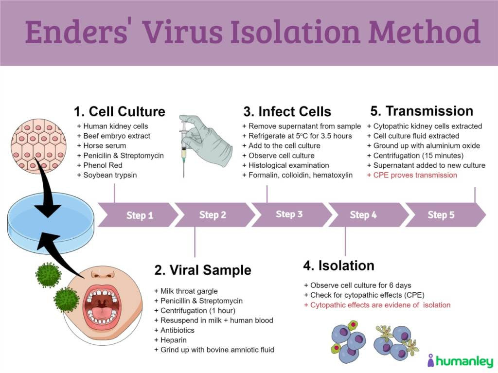
In 1954, John Franklin Enders devised the cell culture experiment as the way to detect whether or not “viruses” are present within the fluids of a sick host. In order for this experiment to work, Enders took throat washings from suspected measles patients (which were obtained in gargled fat-free milk) and added the samples to human and monkey kidney cells. He mixed into the culture bovine amniotic fluid, beef embryo extract, horse serum, antibiotics, soybean trypsin inhibitor, and phenol red as an indicator of cell metabolism. This mixture was then incubated for days and the fluids were passaged (top layer of one culture added to a new culture with a fresh round of additives) on the 4th and 16th days. In doing so, Enders created what he called the cytopathogenic effect (CPE), which is the visual evidence of the death of the cell as it breaks apart and dies from being starved and poisoned. The CPE was claimed to be a specific effect that is used to alert the researchers to the presence of “viruses” in the culture, and by extension, the host.
What is the Cytopathic Effect?
“When a virus invades a host cell, its structure changes. This is known as the cytopathic effect. This condition occurs when the infecting cell causes the lysis of the host cell or when the cell dies due to its inability to reproduce. A virus causing morphological changes in the host cell is known as cytopathogenic.”
https://byjus.com/biology/cytopathic-effect/
However, there is a bit of a problem with using this effect as evidence of a “virus.” For starters, CPE is not specific to “viruses” whatsoever as there are many other factors which are admitted to cause this exact same effect. These include:
Bacteria
Parasites
Amoebas
Chemical Contaminants
Age of the Cell
Incubation Temperature
Length of Incubation
Antibiotics/Antifungals
Environmental stress
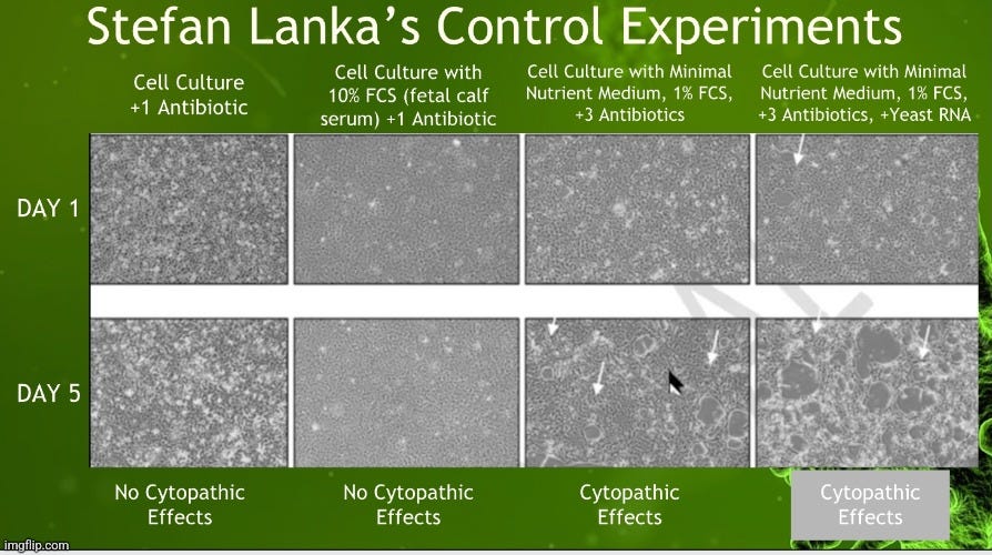
This means that the presence of a “virus” is not necessary in order to explain the observance of CPE. Enders should have known that his experiment was fraudulent when he also observed CPE in his control cultures that did not have any “infectious virus” present. If Enders had any doubt, he would have seen that various researchers in the years following his publication came up with the exact same CPE in their own healthy control cultures, thus shutting the door on CPE being caused by “viruses.” Alas, Enders and the rest of the virology community ignored logic along with these contradictory findings and maintained that this effect was the defining characteristic of the presence of a “virus.”
Beyond the non-specificity of the CPE, there are other reasons that the cell culture experiment is not a valid scientific experiment. These include:
There is no observation of a natural phenomenon and thus, no valid dependent variable.
There is no valid independent variable as there are no purified and isolated “viral” particles confirmed prior to the experiment.
There is no actual valid hypothesis to test against.
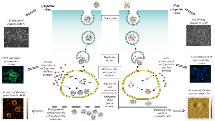
However, even if we were to accept the cell culture as a valid scientific experiment for the proof of the presence of a “virus,” and even if the CPE, which is the basis for the presence of any “virus” within the fluids, was not known to be caused by other factors, this experiment is still mired in falsifiability. This is due to the concept of the non-cytopathogenic “virus.” As the name implies, these are “viruses” that either produce very little or no CPE when cultured. In other words, not all “viruses” are associated with CPE. This caveat allows for virologists to still claim that the cell culture was successful for detecting the presence of a “virus” even when the effect that is supposed to be the result of the “virus” is not observed. On top of that major escape clause, it is allowed for a “virus's” CPE to be different from that seen within the same “viral” family and the CPE pattern may change depending on the type of cell used for culturing:
Cytopathic Effects of Viruses Protocols
“During the time that synthesis of viral components is occurring in the infected cell, the cell undergoes characteristic biochemical and morphological changes. Progression of these changes is most readily observed in cell culture, where infection of cells is more easily synchronized and where the cells can be observed and sampled frequently during the course of infection. Morphological changes in cells caused by viral infection are called cytopathic effects (CPE); the responsible virus is said to be cytopathogenic. The degree of visible damage to cells caused by viral infection varies with type of virus, type of host cells, multiplicity of infection (MOI), and other factors. Some viruses cause very little or no CPE in cells of their natural host. Their presence can be detected visually only by hemadsorption or interference, in which infected cell cultures showing no CPE inhibit the replication of another virus subsequently introduced into the cultures, or in situ by viral antigen or nucleic acid detection. On the other hand, some viruses cause a complete and rapid destruction of the cell monolayer after infection. The microscopic appearance of the CPE caused by some of these cytocidal viruses may be sufficiently characteristic to allow provisional identification of an unknown virus (2).”
“Listed below are several general types of CPE. Keep in mind that a given virus may not conform to the norm for its family, or it may produce different CPE in different host cell types. Characteristic CPE is best viewed by daily observation of cultures that have been infected at a low MOI (<0.1). The best knowledge of viral CPE comes from experience. And control uninfected cells should always be observed to distinguish normal cell changes that occur as cells age from cytopathic effect.”
https://asm.org/ASM/media/Protocol-Images/Cytopathic-Effects-of-Viruses-Protocols.pdf?ext=.pdf
As can be seen, virologists get to have it both ways as the presence or absence of the CPE is not a deal breaker in the identification of a “virus.” Non-cytopathogenic “viruses” are said to shut down proapoptotic mechanisms through the activation of antiapoptotic mechanisms or by inducing alternative transcription programs that preserve the “viral” genome without “virion” production. On the other hand, cytopathogenic “viruses” damage the cell due to high protein expression and “virus” assembly or are actively cytopathic due to expressing proapoptotic molecules. In other words, some “viruses” damage the cells by creating a high amount of “viral” copies while others apparently do not:
Immunogenicity of Cytopathic and Noncytopathic Viral Vectors
“Viruses can be classified as either cytopathic, meaning that cells are killed during the course of infection, or noncytopathic. Some viruses (8) are inherently noncytopathic because their replication programs are relatively benign, while others actively maintain a noncytopathic state by shutting down proapoptotic mechanisms, by activating antiapoptotic mechanisms (7), or by inducing alternative transcription programs that preserve the viral genome without virion production (57). Similarly, some viruses are passively cytopathic as a consequence of a viral replication cycle that damages the cell due to high protein expression and virus assembly or actively cytopathic due to expression of proapoptotic molecules (7, 48).”
https://www.ncbi.nlm.nih.gov/pmc/articles/PMC1488949/
For fun, let's take a quick look at a few of these “viruses” which do not produce CPE. First up is the rabies “virus” which is said to not produce a cytopathogenic effect:
Rabies
“Cultures are infected with cell-culture-adapted MSV and incubated at the appropriate temperature for a defined period. As rabies virus does not normally cause cytopathic effect, this allows several harvests from the same culture.”
While the herpes “virus” is said to produce CPE, according to this study, it seems that virologists get to claim that the “virus” is present even though this effect is not always observed:
Herpes
Non-cytopathic herpes simplex virus type-1 isolated from acyclovir-treated patients with recurrent infections
“Herpes simplex virus (HSV) usually produces cytopathic effect (CPE) within 24-72 h post-infection (P.I.). Clinical isolates from recurrent HSV infections in patients on Acyclovir therapy were collected between 2016 and 2019 and tested in cell cultures for cytopathic effects and further in-depth characterization. Fourteen such isolates did not show any CPE in A549 or Vero cell lines even at 120 h P.I. However, these cultures remained positive for HSV-DNA after several passages. Sequence analysis revealed that the non-CPE isolates were all HSV-1. Analysis of the thymidine kinase gene from the isolates revealed several previously reported and two novel ACV-resistant mutations. Immunofluorescence and Western blot data revealed a low-level expression of the immediate early protein, ICP4. Late proteins like ICP5 or capsid protein, VP16 were almost undetectable in these isolates. AFM imaging revealed that the non-CPE viruses had structural deformities compared to wild-type HSV-1. Our findings suggest that these strains are manifesting an unusual phenomenon of being non-CPE herpesviruses with low level of virus protein expressions over several passages. Probably these HSV-1 isolates are evolving towards a more “cryptic” form to establish chronic infection in the host thereby unraveling yet another strategy of herpesviruses to evade the host immune system.”
https://www.ncbi.nlm.nih.gov/pmc/articles/PMC8789845/
The “norovirus” is special in that it can not be cultured at all. In fact, they don't even have an animal model for the “virus,” only images of unpurified poop particles.
Norovirus
“Noroviruses are classified as members of a category of viruses known as the Calicivirus family. The caliciviruses consist of four groups, of which the noroviruses are the most important human pathogen. Caliciviruses are single-stranded, positive-sense RNA viruses. The caliciviruses have been difficult to study due to their inability to grow in a cell culture system and the lack of a good animal model system.”
Not only does “coronavirus” HKU1 not cause any cytopathogenic effect when cultured, RT-PCR results looking for replication of the “virus” were negative as were attempts to sicken mice with injections of the culture soup into their brains:
Coronavirus HKU1
“After the virus was determined to be a coronavirus, the NPAs were inoculated into RD (human rhabdomyosarcoma), I13.35 (murine macrophage), L929 (murine fibroblast), HRT-18 (colorectal adenocarcinoma), and B95a (marmoset B-lymblastoid) cell lines and mixed neuron-glia culture. No cytopathic effect was observed. Quantitative RT-PCR, using the culture supernatants and cell lysates to monitor the presence of viral replication, also showed negative results. Moreover, intracerebrally inoculated suckling mice remained healthy after 14 days.”
https://viroliegy.com/2021/12/16/woo-coronavirus-hku1-paper-2005/
Finally, we have bovine “viral” diarrhea “virus” (BVDV) which can come in either the cytopathogenic or non-cytopathogenic variety. However, the non-cytopathogenic version is the most prevalent with the cytopathogenic strain said to be rare:
Bovine “Viral” Diarrhoea “Virus”
“Based on the 5′-untranslated region, two BVDV species have been identified: BVDV1 and BVDV2 (Baker, 1995). Each BVDV species is divided into two biotypes, cytopathic (cp) and non-cytopathic (ncp), based on their ability to cause pathogenic effects in cultured cells (Ridpath et al., 2006). The ncp BVDV is the most prevalent biotype in nature and causes acute and persistent infections. In utero infection of cows during the first 120 days of pregnancy with ncp BVDV strains can result in the birth of persistently infected (PI) calves. These PI animals serve as a major source of viral spread in the herd (Brownlie et al., 1989; Darweesh et al., 2015; Polak & Zmudzinski, 2000). In contrast, cp BVDVs are relatively rare; however, when PI calves are superinfected with a cp BVDV strain, they may develop lethal mucosal disease.”
https://onlinelibrary.wiley.com/doi/full/10.1002/vms3.1052
It should be obvious that virologists have created yet another unfalsifiable concept as they allow themselves to claim a “virus” is present whether or not the CPE is ever observed.
Unfalsifiable Concept # 4: “Virus-like” Particles
A small particle that contains certain proteins from the outer coat of a virus. Virus-like particles do not contain any genetic material from the virus and cannot cause an infection.
https://www.cancer.gov/publications/dictionaries/cancer-terms/def/virus-like-particle
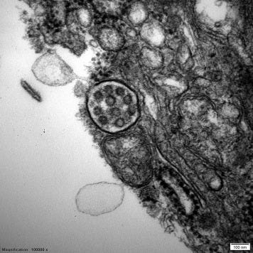
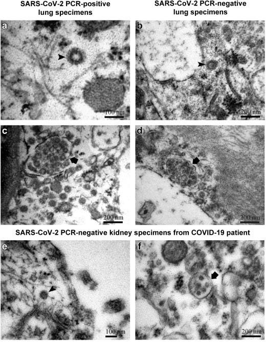
With the rise of “SARS-COV-2,” researchers were seemingly finding the corona particle everywhere they looked in transmission electron microscopy images. Many of these particles were identified in biopsies of tissue sections from “SARS-COV-2” positive patients in an attempt to show that the “virus” was spreading throughout the body and infecting various organs. However, these images were often disputed by other researchers. Instead of the particles being of “SARS-COV-2,” it was argued that these were images of “virus-like” particles (VLP's), such as multivesicular bodies, clathrin-coated vesicles, the rough endoplasmic reticulum, and/or other extracellular vesicles that are normal cellular particles and constituents. According to one paper, there are numerous VLP's that resemble and can be mistaken for “viruses” within the tissues and fluids. Thus, caution was advised when interpreting these images:
Multivesicular bodies mimicking SARS-CoV-2 in patients without COVID-19
“Transmission EM of tissue sections is not a specific or sensitive method for the detection of viral particles; there are numerous structures found by EM that resemble viruses (so-called viral-like particles), such as the well-known endothelial tubuloreticular inclusions (also called myxovirus-like particles). Therefore, caution is suggested when identifying a virus by EM in tissue sections.”
https://www.kidney-international.org/article/S0085-2538(20)30529-9/fulltext
VLP's are not a new thing. They have been described as far back as the 1930's as pointed out in this next article. Unfortunately, much like a “virus,” the definition of just what exactly a VLP is has changed over the decades. It originally meant a particle that looked like a “virus” but was decided, somehow, not to be one. How they were able to determine this without actually purifying and isolating the assumed “viral” particles from the fluids is beyond me. The authors state that not all things that look like “viruses'“ are actual “viruses,” as a VLP can mean either replication-competent structures (“viral” particles) containing a matching viral genome (VLP+) or instead replication-incompetent structures because they lack a “viral” genome (VLP–). This is the equivalent of virologists having their cake and eating it too. The authors attempt to argue that the widespread use of the term VLP from the 1940s–1980s was referring to something that looked like a “virus” as determined by EM, and which might be replication competent (VLP+). In other words, they thought these VLP's were possibly “viruses” but they had no proof that they were replication competent nor had a matching “viral” genome. In the 1990s, VLP was used to describe particles that looked like “viruses” but lacked “viral” genomes and, therefore, were not replication competent (VLP–):
Virus-Like Particle: Evolving Meanings in Different Disciplines
“The phrase “virus-like particle” or VLP has an obvious plainly read meaning: a particle that is like a virus but is not necessarily a virus. There are many particles that resemble viruses, however, and these can differ in properties depending on whether one is looking serologically, through electron microscopy (EM), or using epifluorescence microscopy (EFM). Furthermore, not all things that look like viruses are necessarily actual viruses. As we consider here, VLPs can mean either replication-competent structures (viral particles) containing a matching viral genome (here, VLP+) or instead replication-incompetent structures because they lack a viral genome (here, VLP–). Currently, use of VLP+ (but without the superscript) is widespread among viral ecologists. Use of VLP– (but also without the superscript) is widespread among biotechnologists developing vaccines and gene delivery systems. How did this divergence in meaning of VLP come into being?
VLPs as Potentially Replicative Viruses in Medical Science (VLP+)
The earliest use of “virus-like particle” was in a 1933 article by Macfarlane Burnet, who was studying the neutralization of bacteriophages by antiphage serum.1 VLP was seen again in 1939 by Eagles and Bradley to describe agglutinations from exudates mixed with serum from patients recovering from rheumatic fever.2
Beginning in the 1940s and 1950s, there was a dramatic increase in use of VLP (or the similar, “virus-like bodies”), generally equivalent in meaning to VLP+, as researchers began using EM to study animal cell ultrastructure. Many of the reports of particles in cells that looked like EM images of animal viruses were from studies of cancer cells3,4 or from cell lines derived from tumors.5 VLPs were also seen in cells infected with known viruses.6,7 Figure 1A shows an EM typical of this usage of VLP. Some researchers discussed whether VLPs were viruses (e.g., Fawcett8). Others concluded that VLPs were indeed viruses and used “VLP” to describe what was seen in electron micrographs but “virus” when not talking about micrographs.7,9
In the 1950s–1970s, VLPs were seen in EM images from noncancer systems such as virus-infected plant cells,10,11 mushrooms,12 in the milk from cows in herds with lymphosarcomas,13 yeast infected with P1 virus,14 and human liver cells from persons infected with hepatitis.15 This last article, and others working with hepatitis, also associated VLPs as well as genuine viruses with disease-specific antigens.16,17 With the variety of VLPs being seen, Bernhard proposed a classification system for VLPs based on size and morphology and this became commonly used.18,19 Although often not discussed in these articles, these VLPs potentially were viruses with nucleic acid cores, that is, as equivalent to VLP+, although only partially assembled capsids were seen as well.”
Conclusions
“To summarize, widespread use of the term VLP from the 1940s–1980s was referring to something that looked like a virus as determined by EM, and which might be replication competent (VLP+). In the 1990s, however, VLP came instead to describe particles that looked like viruses but lacked viral genomes and, therefore, were not replication competent (VLP–). These VLP– constructs retain many physical characteristics of a virus, including immunogenic properties, allowing them to be used as vaccines. Also in the 1990s, an additional usage of VLP was introduced, one that is equivalent to VLP+. These are particles that are like viruses because they are small and contain nucleic acid. They thereby can fluoresce when stained with a nucleic acid-binding dye. Such VLPs can be used to approximate virus prevalence in environments without the need for either complicated EM observations or culturing.
These different meanings of VLP appear to be used by distinctly different subdisciplines of biology. During interdisciplinary discussions, such as during presentations at general meetings, the existence of multiple definitions for a single term can be confusing. We hope, therefore, that this commentary will remind researchers that VLP now has multiple somewhat distinct meanings. Furthermore, when ambiguity is a concern, as an aid toward distinguishing VLP as employed in viral ecology from that used in biotechnology or vaccine development, we suggest use of the construct, environmental VLP.”
https://www.ncbi.nlm.nih.gov/pmc/articles/PMC9041479/
We can see that the current definition of VLP in use today lines up with particles that are indistinguishable from “viruses” that do not contain a “viral” genome.
“Virus-like particles (VLPs) are protein complexes that are similar to, or even indistinguishable from, native virus particles, but that do not contain the viral genome. Hence, they cannot replicate but can mimic the viral antigenicity without being pathogenic. Depending on the biology of the virus, VLPs can be icosahedral or helical (rod-shaped), consist of one or more capsid proteins in multiple copies and may be enveloped or not. While VLPs can occur naturally (e.g., poliovirus empty capsids outside of the cell), they can also be recombinantly produced by expressing the proteins required for VLP production.”
https://www.sciencedirect.com/science/article/pii/B9780128145159000679
This allowance for the existence of “virus-like” particles is yet another way where virologists get to have it both ways. If the particles claimed to be “viruses” are found within tissues where they shouldn't be or in “uninfected” individuals, virologists can easily claim that these are just VLP's and are not actually the “virus.”
This has given virology quite the escape clause as there are many particles that are said to look exactly like, and are regularly mistaken for, “viruses” that are found within both healthy and unhealthy individuals.
If the particles claimed to be “viruses” are able to be found within both the sick and the healthy and can then be said to be either a replication-competent “virus” or just particles that look like one based on a whim, virology has given itself yet another way of making their theory entirely unfalsifiable.
Virology’s Unfalsifiable Hypothesis
By looking at the examples of unfalsifiable concepts present in virology, you should be able to see how variable and vague the concepts of virology truly are. As stated by Oxford Reference, pseudoscience “provides no room for challenge and tends to dismiss contradictory evidence or to selectively decide what evidence to accept.” They conclude that pseudoscience is “nothing more than a claim, belief, or opinion that is falsely presented as a valid scientific theory or fact.” It is clear that virology fits this pattern and is, by definition, pseudoscience. Virologists have come up with various ways in which they can attempt to explain away contradictory evidence and findings that should have rightly falsified their hypothesis. Instead, virology seeks to confirm the theory rather than challenge it, which is a hallmark of pseudoscience:
Drawing the line between science and pseudo-science.
“The big difference Popper identifies between science and pseudo-science is a difference in attitude. While a pseudo-science is set up to look for evidence that supports its claims, Popper says, a science is set up to challenge its claims and look for evidence that might prove it false. In other words, pseudo-science seeks confirmations and science seeks falsifications.
There is a corresponding difference that Popper sees in the form of the claims made by sciences and pseudo-sciences: Scientific claims are falsifiable -- that is, they are claims where you could set out what observable outcomes would be impossible if the claim were true -- while pseudo-scientific claims fit with any imaginable set of observable outcomes. What this means is that you could do a test that shows a scientific claim to be false, but no conceivable test could show a pseudo-scientific claim to be false. Sciences are testable, pseudo-sciences are not.”
Virology allows for:
“Viruses” to be found within the sick and also within the healthy.
Theoretical antibodies to be found in the “infected” and not found in the “infected,” while being either a sign of “protection” or a sign of chronic disease.
The presence of the “virus” within the cell culture determined by both the observation of the cytopathogenic effect as well as the lack of the observation of this efffect.
The same particles seen in EM images claimed to be either pathogenic “viruses” or non-pathogenic “virus-like” particles.
It is clear that there is no way to be able to falsify the “viral” hypothesis when contradictory concepts are ultimately allowed to coexist in order to explain away inconvenient findings. The “viral” theory is then reworked to allow for the incorporation of the contradictory findings to further confirm and support the unfalsifiable premise. Germ theory and virology are a circular system devoid of logic and reason that have been deceiving humanity for the last two centuries. Isn't it far past time to demand that they show how their hypotheses and theories are falsifiable?
examined the issues related to most of the published "scientific" research being false. clarified some misconceptions about Dr. Stefan Lanka's control experiments. took a look at a 2017 WHO document that "anticipated" the need for all of the world to establish virology labs.








Your excellent post is another well-sourced take-down of the LIE from you.
I am exhausted that some people still want to boost and mask. After all the evidence that has emerged, it is now at the level of superstition.
Been following quietly for a while now. I want to contribute an article of my own to sum all of this into an easily digestible form. I'm hoping to upgrade to paid when my next pay packet comes in. Cheers.