Imagine for a moment that you had no concept of Santa Claus. You have never heard of the jolly fat man in the red jumpsuit who delivers presents to all of the good boys and girls around the world. You wake up one morning, walk downstairs, and you discover presents under a beautifully decorated tree in the living room along with stockings hung on the fireplace mantel filled with wonderful goodies. There are traces of soot and some faint footprints on the carpet leading from the fireplace to the tree. Sitting on the stand next to the rocking chair is a plate with leftover cookie crumbs and a half drank cup of milk. There is a hand-written note thanking your family for the delicious snack signed by someone named Santa. While cautiously excited at first, you begin to panic, fearing that a stranger has broken into your home while you slept. However, instead of taking anything of value, this stranger left you with more than you had before. Rather than cover up the crime, this Santa character left plenty of evidence implicating himself in this holiday breaking-and-entering caper.
You run to your parents, feeling somewhat bewildered and stunned as there is no sign of any forced entry into the house. Filled with a sense of concern for the safety of your family, you explain to them every last detail of the perplexing scene that you stumbled upon. Your mother lets out a little laugh, gives you a gentle hug and a kiss on the forehead in order to calm you down, and tells you to relax.
“Everything is OK. It was just Santa Claus.”
Santa Claus?!? Confused by your parents seeming disregard over the fact that there was an intruder in the house, you ask them how they can remain so calm knowing that this Santa Claus broke in while everyone slept. You press them for more details.
Over the course of the next few minutes, you are told a fantastical tale about a hefty old, bearded man who possesses superhuman abilities. This senior citizen, with no concern over developing diabetes from his cookie binging ways, is somehow able to fly around the entire world.in a single night on a sleigh of eight magical reindeer. He is able to transport himself up and down through the tightest of chimneys with ease. Within a giant burlap sack that he lugs around with him everywhere is an endless supply of just the right gift-wrapped toys made by a village of elves that he lives with in the North Pole. Amazingly, this elderly man knows exactly who to give the gifts to without question as he watches the children of the world all year round in order to make sure whether they have been naughty or nice.
After this wild explanation by your parents attempting to unravel the mysteries of the marvelous scene that you witnessed downstairs, you are left in a state of utter shock. There are numerous questions running through your head. You take a moment, collect yourself, and then ask the most logical one that comes to mind:
“How do you know that this Santa Claus actually exists and travels across the world on a sleigh of eight flying reindeer delivering presents to all of the good boys and girls?”
Taken aback by your simple question, your parents look at each other for a moment, and with a slight smirk, your father responds.
“Didn't you see the presents left under the tree?”
You explain that, of course you saw the presents downstairs, but how are you supposed to know that the presents were placed there by this Santa Claus person? Couldn't the presents have been left by someone else, perhaps even by your parents themselves? Your mother starts to look a bit nervous.
“You saw the cookie crumbs and the half-drank cup of milk, right?”
Again, you explain that this could easily have been the act of either of your parents. How do cookie crumbs and a glass of milk equal a magical fat man? Your father, getting a bit upset, responds back.
“What about the hand-written note thanking us for the delicious snack?”
To this, you reply that the handwriting looked suspiciously like your mother's, which elicits a faint gasp from her. Your mother shoots back.
“But what about the footprints and the soot left by the fireplace?”
Your parents seem fairly content that this is solid evidence of a fat man who can magically transport himself down and back up the chimney with a giant bag of toys. But alas, you explain that they could have easily made the footprints themselves with a pair of boots and some fireplace ash. Your parents are now sweating a bit. Their explanations are not logical, and they realize that they are failing to convince you. However, in a last desperate attempt to convince you that jolly ol’ Saint Nick is a real person, your father pulls out his smart phone with an image of Santa Claus caught sneaking around inside your house.
“Look! Here is an image of Santa delivering the presents to us! We caught him right in the act with our security camera last night.”
You sit down for a moment, and your parents start to feel a sense of relief. That is, until you ask them to show you when, where, and how the image was obtained from the security camera. Your father, clearly flustered now, simply refuses to respond any more about the photo or any other questions that you have that further poke holes into their story. Your mother throws up her hands in frustration and says something about not raising a child who disbelieves in Santa. She lashes out:
“All of the children believe that Santa Claus exists, and all of the evidence points to him existing! Are you denying that Santa is real?”
You counter that all of the evidence that your parents presented can be explained away. The simplest explanation is that they were the ones who set up the scene, and they were the ones who created the evidence implicating a supernatural being. All of the evidence that they offered did not present a clear case that Santa existed, nor that this man was capable of the amazing qualities that they had attributed to him. All your parents had was evidence that, if believed, could lead one to possibly buy into the story that such a person existed. This is why there are so many children who are willing to believe in this fantastical being without question as they do not look at this evidence critically and logically. However, what your parents failed to present when making their case was any evidence that proved Santa Claus existed that did not rely of the necessity of inference in order to come to such a conclusion.
This little exercise in creative writing is meant as a metaphor for the interactions many of us face regularly when we discuss the lack of scientific evidence supporting the existence of pathogenic “viruses” with those who believe in the germ theory story. Sadly, these conversations are rarely as nice as the one I presented in the above Santa Claus scenario. However, they always follow the same pattern where those who defend the story supply plenty of circumstantial evidence to us that is supposed to stand in as proof for the existence of the fictional entities. They give us presents under the tree, cookie crumbs and glasses of half drank milk, dirty footprints, images of someone dressed as Santa, etc. However, what they can never do is show that Santa is a real individual rather than someone dressed up and made to look like the real deal. They can never demonstrate that the person playing the part of Santa possesses all of the magical qualities attributed to him. They can never establish that Santa actually lives in a village at the North Pole. All they present is evidence that, when examined critically and logically, can be explained away as being created by the very people who are attempting to sell us on the idea that such a person as Santa Claus actually exists in the first place.
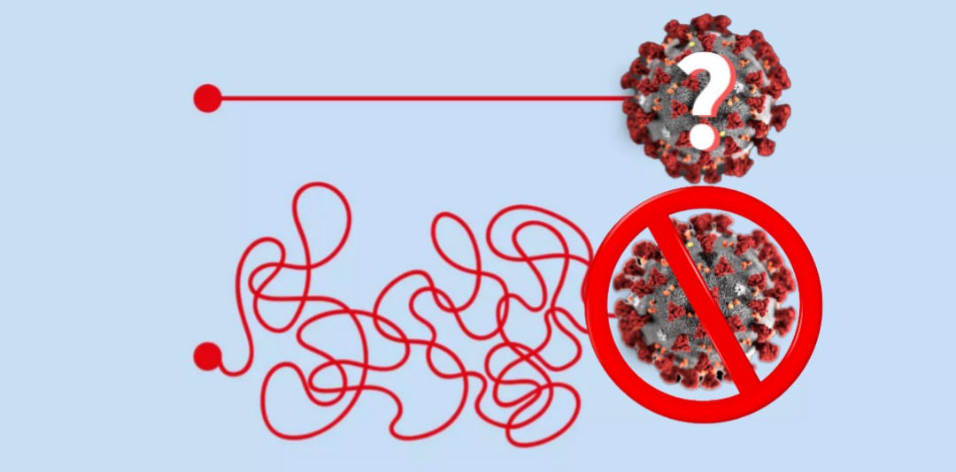
What this ultimately boils down to is virologists attempting to make the case that a “viral” culprit committed a crime using only circumstantial, or indirect, evidence. However, they are attempting to do so while offering no direct evidence that the culprit even exists at all. They are blaming a defendant who isn't even in the court room. There is big difference between providing indirect evidence over direct evidence, and it is a major sticking point that keeps many smart people mistakenly believing in fairy tales. Thus, I want to examine the difference between both types of evidence and show why the six most common forms of indirect evidence that virology supplies, beyond being unscientific, is not adequate proof for the existence of the invisible pathogenic entities.
“This type of functional correlation is the only way in which the behavior of viruses may be studied. Since viruses cannot be seen directly, some indirect evidence of their presence must be forthcoming. This indirect evidence is the effect the viruses produce in animals. If no experimental animal is susceptible, then there is no way of observing the presence or absence of the virus, let alone its functional correlates with other states or conditions. Advance in the study of a virus disease is usually dependent on the discovery of a susceptible host. If that host happens approximately to reproduce the original disease, well and good. But such reproduction is not essential.”
-Lester S. King
https://doi.org/10.1093/jhmas/VII.4.350
According to Lester S. King's 1952 paper Dr. Koch's Postulates, virology has a bit of a direct evidence problem as “viruses” cannot be seen, and thus, there is no direct evidence confirming the existence of these invisible entities. This sentiment was reinforced in 2006 with the 6th edition of Introduction to Modern Virology where it is stated that “Viruses occur universally, but they can only be detected indirectly.” A 2020 review on the history of early bacteriophage and virology research admitted that the indirect evidence collected would not have met the modern quality standards of direct evidence and visualization, thus it would fail to convince the peer reviewers of today. Nevertheless, the evidence that would not pass muster today was used to create the concept of the “virus” as a transmissive genetic program:
“While the quality standards of modern science impose obtaining direct evidence and visualization of the phenomena and objects studied, the methodology of science of the earlier period largely relied on making conclusions based on multistep indirect deductive reasonings, which would not be accepted as sufficiently convincing by the peer reviewers of the modern scientific journals. Nevertheless, these were the approaches that required extensive laboratory work and enormous intellectual efforts in planning the experiments and analyzing the obtained data, and as a result of these approaches, main postulates of molecular biology and the highly important biology concepts were developed including the concept of a virus as a transmissive genetic program.”
Thus, it is not a stretch to say that in order to convince themselves that these entities exist, virologists only offer up indirect evidence that is used to convey the presence of these entities.
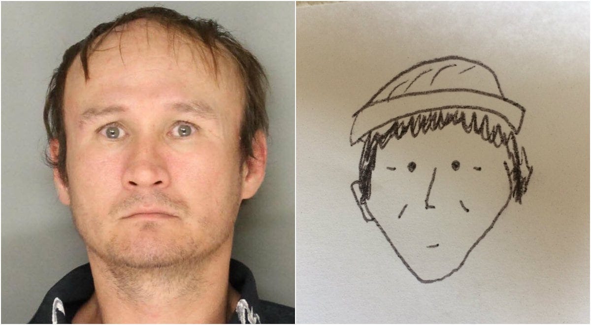
For clarification, the law defines indirect evidence as a combination of facts that, if true, permits a reasonable person to infer the fact. This type of evidence does not directly prove the matter at issue. On the other hand, direct evidence directly demonstrates a fundamental fact, and it is evidence that directly proves the matter. While both types of evidence are permittable and the law makes no distinction as to putting more weight on one versus the other, the indirect evidence must be strong enough and convincing enough to make the case. However, no matter how much indirect evidence is amassed in order to claim a defendant is guilty of a crime, no prosecutor can try a defendant who does not actually exist. A prosecutor cannot lay out indirect evidence before a jury for them to infer the defendant into existence. The defendant must actually be present and accounted for in order for the evidence to matter. This is the biggest problem for virology as it does not have any defendant. All that is presented is the equivalent of a bad police sketch.
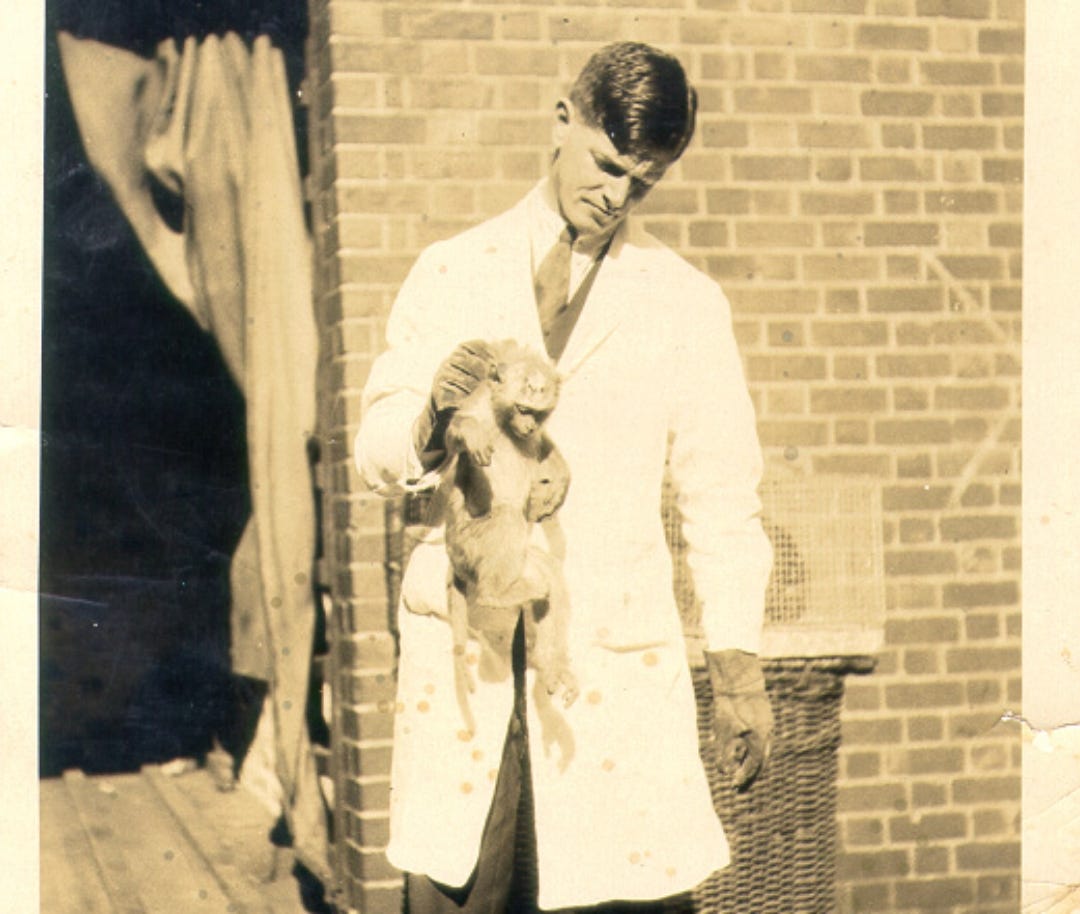
The idea of a “virus” existed long before any submicroscopic particles were ever claimed to be one. King pointed out that, as “viruses” could not be directly observed, the only way for a virologist to study a “virus” was to find a host animal that they could sicken experimentally. Without one, they had no ability to study a “virus” or to determine whether a “virus” was actually present. According to Field's Virology textbook, as early virologists were unable to see their agents in a light microscope and were often confused by the great diversity of the effects seen, they had to rely on faith that they were actually working with the assumed “viruses” in their studies. Without the ability to see these entities, virologists relied upon the size of the pores of a filter that were too small for known bacteria to pass through in order to claim that a “virus” was able to do so. They used physical and chemical reactions to alcohol and ether to claim that their entity was present. Virologists relied on the emerging field of the equally invisible antibodies to claim that non-specific results from combining blood samples were indicative that a specific antibody was reacting to a specific “virus.” They would perform grotesque animal experiments in order to try and recreate disease, such as grinding up the brain and spinal cord of a deceased 9-year-old with other substances in order to inject this soup into the brains of monkeys to create paralysis, as seen with polio. These are all forms of creating effects experimentally without an identified cause in order to assume the presence of the unidentified cause indirectly.
Until John Franklin Enders established the fraudulent cell culture technique in 1954, virology was running on fumes. They could not purify (free the sample of host materials, contaminants, impurities, etc.) and isolate (separate from everything else) the particles claimed to be “viruses” directly from the fluids of a sick human or animal. They could not obtain any images of just the purified and isolated “viral” particles. They could not prove that they could recreate the exact disease via natural routes of exposure with the fluids of a sick human or animal. In fact, researchers failed repeatedly to do so. They could not provide any evidence supporting virology that was derived from the scientific method as they had never actually directly established that their invisible entity existed anywhere in nature or within the fluids of a host. Thus, all that the virologists could do was rely of the creation of indirect evidence and pretend that, cumulatively, this was irrefutable proof supporting the idea of pathogenic “viruses.” As time passed, researchers in various fields continued to establish even more indirect detection methods in order to pile on top of the stack of pseudoscientific evidence already collected. Let's explore the six most common forms of indirect evidence that virologists have utilized so that we can gain an understanding as to how they tricked themselves and the rest of the world that their fictional concept actually exists.
Indirect Evidence #1: Filterability
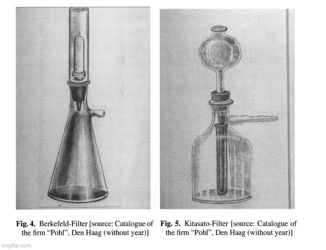
In a recent article, I detailed how the idea of the “virus” was dreamt up when bacteriologists could not identify a bacterial cause in many cases of disease. These researchers were unable to fulfill Koch's Postulates, the logical criteria established by German bacteriologist Robert Koch in 1890 that were deemed necessary to fulfill in order to claim any microbe as a causative agent of disease. They therefore needed an explanation for any experimental diseases that they were inflicting on animals and plants. As the fluids used in these experiments were claimed to still be able to cause disease when filtered through Chamberland filters said to keep out all known bacteria, it was assumed that something smaller than a bacteria must have passed through in order to cause the symptoms of disease. Granted, the original concept was either that of a poison or an even smaller bacterium. The “virus” as we know it today, i.e. an obligate replication-competent intracellular parasite surrounded by a protein coat, did not present itself until the 1950s.
Going back to Field's Virology, it is stated that these “filterable viruses” were classified as distinct based solely upon the pore size of the filter. The only way that one “virus” could be distinguished from another was by the symptoms of disease inflicted, which as I pointed out in my article linked above, were most likely the result of the experimental methods and unnatural means of inoculation utilized, such as wounding the hosts in various ways, rather than the presence of any “virus:”
“In the early 1900s, viruses were initially classified as distinct from other organisms simply by virtue of their ability to pass through unglazed porcelain filters known to retain the smallest of bacteria. As increasing numbers of filterable agents became recognized, they were distinguished from each other by the only measurable properties available, namely the disease or symptoms caused in an infected organism. Thus animal viruses that caused liver pathology were grouped together as hepatitis viruses, and viruses that caused mottling in plants were grouped together as mosaic viruses.”
According to Alfred Grafe's A History of Experimental Virology, the small size meant that “viruses” remained macro- and microscopically undetected. Depending on the pore size and quality of the filter used, “viruses” were either filterable, or they were not. There were issues with adsorption properties of the filters utilized leading to a solid film sticking to the surface. Colloids, which include gels, sols, and emulsions that cannot be separated out by ordinary filtering or centrifuging, appeared as proteins in the solutions that were to be filtered. In other words, these were not purified solutions of nothing but “viral” particles. In order for a “successful” filtration, the pore size had to be calculable, adsorption had to be excluded, and the organisms had to be round. However, as researchers were using filters meant for dialysis, these requirements were not met, meaning that to ensure accurate results, improvements were necessary. This should lead anyone to wonder how accurate the early research was with the invisible “filterable viruses.” However, these issues did not stop virologists from claiming the discovery of numerous such invisible entities, and by 1937, they had firm estimates of the particle size for 23 "filterable" human and animal pathogens, 13 types of bacteriophage, and TMV with sizes ranging from 8 to 230 nm. In other words, the researchers had decided on sizes for the “viruses” that they could not see based upon the pore size of the filters used:
“Filterable viruses were distinguished by negative biological and physical features: It was not possible to culture them, and because of their small size they remained macro- and microscopically undetected. They were or were not filterable, depending on the pore size and quality of the filter used. The range of physical methods employed during the first three decades of the 20th century to characterize viruses included filtration, centrifugation, adsorption, electrophoresis, and optical methods.
In the early stages, the same filter candles or filter layers used in experiments with bacteria were applied to filterable viruses. Whilst bacteria, as a result of their size, did not pass through intact filters, whose greatest drawbacks were clogged pores and germ-infected cracks, for the smaller, filterable viruses, new problems arose. Here, filtration results depended both upon the average and maximum pore size of the filter. In addition, there were annoying adsorption properties of the filters, and colloids which appeared, for example, as proteins in the solutions to be filtered.
The aims of filtering were twofold: to isolate the pathogen and to obtain the most accurate information possible about the organism. Both aims were achievable providing three prerequisites were fulfilled: the range of pore size about the mean had to be calculable; adsorption by the filter had to be excluded or measurable; and the organisms had to be as round as possible to enhance permeability. Since the filters in use at the time in bacteriology and in colloid chemistry had been developed in part for the purpose of dialysis, they did not necessarily fulfil these basic requirements. Their improvement or further development was, therefore, essential.”
“The list of filterable viruses in 1915 - the year the bacteriophage was discovered - totalled 34, related to human, animal and plant diseases. By 1937 scientists had firm estimates of particle size for 23 "filterable" human and animal pathogens, 13 types of bacteriophage, and TMV. The particle sizes ranged from 8 to 230 nm. The smallest known germs, besides the bacteriophages, were the pathogens for poliomyelitis, yellow fever, encephalitis, FMD and TMV, ranging in size from 8-30 nm. Despite significant improvements in filtration techniques, the process continued to be problematic. The wide variation in pore diameter persisted, producing unreliable estimates of particle size. Isolating pathogens was connected with a high rate of germ loss because of adsorption by the filters, the adsorbing properties of filterable viruses known as biocolloids, and extraneous deposits on the filters themselves.”
The “virus” was nothing but a guesstimate. As noted by the authors of Introduction to Modern Virology, the first definition for “viruses” was presented entirely in negative terms as these entities could not be seen and could not be cultivated. In other words, the “viruses” were nowhere to be found. Thus, the most important evidence used for the creation of the “virus” concept which was used to justify their existence was the indirect evidence of nothing being retained by bacteria-proof filters.
Indirect Evidence #2: Plant & Animal Experiments
“The only possibility of providing a direct proof that comma bacilli cause cholera is by animal experiments. One should show that cholera can be generated experimentally by comma bacilli.”
-Robert Koch
Koch, R. (1987f). Lecture cholera question [1884]. In Essays of Robert Koch. Praeger.
Robert Koch considered the only direct proof that a microorganism could cause a disease was by experimenting with a pure culture of the microorganism, exposing a susceptible host to it, and recreating the exact same disease. However, it was a very difficult task trying to recreate the exact same symptoms in animals as that seen in humans. In fact, in many instances, it was considered impossible. This was admitted in Alfred Grafe's “A History of Experimental Virology:”
“Since Koch knew, after his 1884 experience with cholera, that it was often impossible to induce a disease experimentally in animals and yet not harbour the slightest doubt about the germ theory, he augmented these guidelines.”
Thus, Koch essentially abandoned the only direct proof that he stated investigators could offer. Researchers have been attempting to excuse this difficulty of recreating the same disease ever since. Lester King was very critical of the requirement to recreate the disease as seen in humans exactly in animals. He stated that the attempts to do so, such as in the case with yellow fever, were examples of “the logical fallacy of begging the question on a truly colossal scale.” This fallacy involves an argument where the conclusion is assumed in one of the premises. In this instance, King pointed out that investigators took a patient claimed to have yellow fever, recovered an agent that they assumed was the cause, injected this agent into guinea pigs which expressed similar but not identical symptoms to the patient, and in a case of circular reasoning, the agent was assumed to be the cause of the human condition based upon the symptoms seen in guinea pigs.
“Yellow fever, perhaps, is a good example of the logical fallacy of begging the question on a truly colossal scale. Investigators have started with a patient whom they call an example of the disease under study. From this patient they recover some microbiological agent whose etiological connection is assumed. When injected into animals, the agent produces disease symptoms suggestive of the original condition. But obviously, a disease in guinea pigs is not identical with a disease in humans in spite of any similarities. Now comes the circular reasoning. The recovered organism is assumed to be the cause of the human condition. In proof, the investigator points to the disease produced in guinea pigs. But how do you know that the disease in guinea pigs is fundamentally or essentially similar to the disease in humans? Because it is produced by the same agent which "causes" the disease in humans. This reasoning which blandly assumes what requires to be proven is the type of reasoning indulged in, for example, by Sanarelli. It is clearly invalid. For cogency in demonstration further extensive evidence must be forthcoming.”
Being able to experimentally produce symptoms of disease in animals that were similar but not identical to the human disease was not considered ultimate proof by King that the agent used was, in fact, the causative agent of the human disease. He argued that further proof was required in that the fluids from the diseased patient must be shown to be clearly “infectious” first before searching for possible etiological agents within the fluids. All potential organisms must be identified from the fluids, and then discarded by a process of elimination:
“Examination of Koch's original paper shows that he did not in any way demand the duplication of the original disease. To interpret Koch's postulates we must start, first of all, with a disease which is proven to be infectious. We must also be aware that some crude infectious material (i.e., blood, sputum, feces, necrotic tissue) is capable of transmitting the infection. Koch's postulates come into play in determining exactly what precise agent is the responsible factor within the crude infectious material. If, for example, we show that pneumonia is infectious and that the sputum carries the infection, then we can apply Koch's postulates to the sputum to find out which organisms are contaminants and which, if any, is the "etiologic agent.”
Let us say that we determine the infectiousness of the sputum by injecting it into mice. Then we could culture the sputum and recover, say, five different organisms, each of which is propagated in pure culture. By injecting each of these into mice we might find that four are harmless, but that the fifth always kills the mouse. By Koch's postulates, according to Koch's own words, the fifth culture could be considered the etiology. Koch specifically states, as quoted above in reference to tuberculosis, that the isolated bacteria must produce "the same clinical picture .. . as is obtained empirically by the injection of naturally developed" material.”
Obviously, as “viruses” are said to be invisible and unculturable, this process of elimination does not work beyond claiming that no bacteria are left after filtration, and that there must be some other entity remaining that can produce disease hiding within the filtrate. However, without being able to see what is in the filtrate, there could be multiple potential causative agents residing amongst each other. These filtrates were then used to experimentally create disease in plants and animals in the late 1800s and beyond in order to indirectly claim a “viral” cause that could not be identified. However, the methods used to “infect” healthy hosts were anything but natural, and the filtrates used were anything but pure.
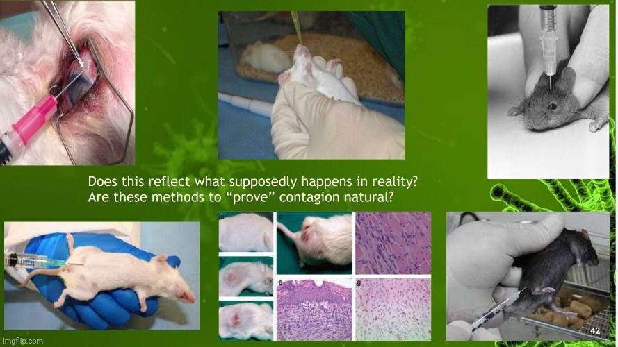
Here are a few examples of how researchers created disease experimentally:
For Tobacco Mosaic “Virus” (TMV), Martinus Beijerinck crushed up diseased plant tissues with other chemicals, including highly toxic formalin, and injected the plant with a syringe. In order to speed up disease, a deeper wound was created with a knife and more material was used. The disease was not identical to that seen in nature.
For Foot-and-Mouth Disease (FMD), Friedrich Loffler scarified the lips, mouths, and mucous membranes of animals and rubbed diseased materials into the wounds, claiming any resulting blisters were a successful transfer of disease.
For Rabies, Louis Pasteur emulsified diseased dog brains with other ingredients and injected this into the brain of other dogs in order to create neurological disease and death.
For Polio, Karl Landsteiner and Erwin Popper took a diseased piece of spinal marrow from a nine-year-old boy, chopped it up, dissolved it in water and injected one or two whole cups intraperitoneally (into the abdominal cavities) of two test monkeys. Simon Flexner and Paul Lewis experimented with a similar mixture and injected this into monkeys’ brains. When paralysis and death occurred, these researchers claimed success.
For herpes simplex, investigators in all parts of the world used fluids inoculated into the scarified cornea of rabbits to produce a vesicular eruption followed by an intense keratoconjunctivitis which was considered a successful recreation of the disease. In other words, damaging the eye and inoculating fluids into it caused swelling and redness.
For influenza A, Thomas Francis Jr.
took specimens of sputum in 50 percent glycerine, which were separated from the glycerine, washed and emulsified in Locke’s solution. The material was inoculated intranasally into ferrets under ether anesthesia. One animal with a high temperature was sacrificed on the third day after inoculation; the lungs and turbinates were removed and ground with sand and meat infusion broth. Two animals were then inoculated intranasally with unfiltered suspensions and one with a Berkefeld V filtrate of the material.
There are many more grotesque examples of how researchers attempted to recreate diseases experimentally. There is absolutely nothing natural about this process. All that virologists are doing is torturing plants and animals, wounding and injecting them in unusual ways with unpurified solutions and claiming that the resulting symptoms are proof that they were working with the invisible causative agent. More often than not, proper controls were not carried out that would have shown that the cruel experimental methods, and not the filtrates, were the cause of the symptoms of disease. No wonder King was adamant that this was not direct proof of cause.
Indirect Evidence #3: Antibodies
"An indirect way of visualising viruses is to use antibodies (much like the ones your body makes in response to infection) to tag viruses with fluorescent molecules that give off light when they absorb certain types of radiation.”
https://theconversation.com/five-techniques-were-using-to-uncover-the-secrets-of-viruses-144363
In Chapter 1 of Grafe's A History of Experimental Virology discussing the emergence of experimental virology, it states that early researchers assumed, i.e. suppose to be the case without proof, that there were substances within the blood and the body fluids that targeted invasive pathogens.
“As for humoral immunity, it was assumed that there were substances in the blood and in the body fluids which could attack and destroy invasive microorganisms.”
What these substances were was unknown as, just like “viruses,” they were too small to be seen by light microscopy and could not be properly purified and isolated directly from the fluids in order to be studied. The idea that these substances, which ultimately became known as antibodies, were in the blood was brought about by the work of Emil Von Behring and Shibasaburo Kitasato in 1890. In order to come to this conclusion, the researchers pretreated animals with iodine trichloride and then injected unknown culture and/or sera into the stomach of these animals. They then took the blood from the pretreated animals and injected that into the stomach of another pretreated animal. When the second animal did not become sick, it was assumed that the first animal had developed antibodies which were conferred to the second animal. Or at least that was what Von Behring claimed. His partner, Kitasato, published his own articles stating that he believed that what they had observed was a standard reaction known as habituation to poison. In other words, the body gradually became more efficient at removing the toxins and built up a tolerance, as seen with alcoholics and drug addicts after repeated exposures.
As the antibodies are invisible, they are nothing but an imaginary concept based upon indirect experimental effects just as their “virus” counterparts are. Despite this fact, the researchers decided that these entities existed based upon the reactions observed in animal experiments and the mixing of samples of blood. It was then decided to conceptualize how these substances look, form, and function, which fell to German physician Paul Ehrlich in the late 1800s. Ehrlich is credited with coming up with the name antibodies in 1891, as well as establishing the antigen-antibody relationship and the lock-and-key mechanism in his Side-Chain theory of antibody production that was developed in 1897 and presented in 1900.
However, there were many who disagreed with Ehrlich's work and there was much doubt about the make-up and even the existence of these entities. Felix Le Dantec, who denied the physical existence, stated that Ehrlich “added nothing to the explanation of the imaginary invalid.” Henry Dean, professor of Pathology at the University of Cambridge and Master of Trinity Hall, Cambridge, noted that “agglutinins, precipitins, amboceptors are mere words, and a passive belief in the existence of such bodies tends to impede rather than advance our understanding of what is actually taking place;” while adding that “ignorance, however aptly veiled in an attractive terminology, remains ignorance.” In a 1902 letter nominating Ehrlich for the Nobel Prize, Bernhard Naunyn stated that “Ehrlich’s contribution should still be regarded as tentative or premature, since the isolation and purification of the relevant substances (namely, “antibodies”) would long remain a chimera.” Julius Citron’s 1910 statement in a German textbook admitted that antibodies had never been isolated and that their status as a chemical entity remained uncertain:
“In order to learn the nature of these antibodies attempts have been made to isolate them chemically. Thus far all such trials have been unsuccessful. It is even uncertain whether these so-called antibodies are definite chemical entities. Only the effects of the serum as a whole are known, and the ingredients in it to which these activities are attributed are thought of as antibodies.”
-Julius Citron
This statement was backed up in 1929 by H.G. Wells in his book The Chemical Aspects of Immunology:
“We attribute this altered reactivity [of sera] to the presence of “antibodies,” despite the fact that we have absolutely no knowledge of what these antibodies may be, or even that they exist as material objects. Like the enzymes, we recognize them by what they do without discovering just what they are.”
https://archive.org/details/chemicalaspectso0000hgid/page/4/mode/1up?q=Attribute
Thus, we have a similar situation with antibodies that we see with “viruses” where unproven invisible entities are claimed to exist based upon indirect evidence of effects observed in a lab. The researchers claim that if they mix together blood samples and get a reaction, this is evidence that not only that the antibody exists, but so, too, does the “virus.” An example of this is the complement fixation test, which is where a mixture of sheep blood cells, guinea pig serum, and a patient sample are combined together in order to observe a reaction:
“The complement fixation test consists of two components. The first component is an indicator system that uses combination of sheep red blood cells, complement-fixing antibody such as immunoglobulin G produced against the sheep red blood cells and an exogenous source of complement usually guinea pig serum. When these elements are mixed in optimum conditions, the anti-sheep antibody binds on the surface of red blood cells. Complement subsequently binds to this antigen -antibody complex formed and will cause the red blood cells to lyse.
The second component is a known antigen and patient serum added to a suspension of sheep red blood cells in addition to complement. These two components of the complement fixation method are tested in sequence. Patient serum is first added to the known antigen, and complement is added to the solution. If the serum contains antibody to the antigen, the resulting antigen-antibody complexes will bind all of the complement. Sheep red blood cells and the anti-sheep antibody are then added. If complement has not been bound by an antigen-antibody complex formed from the patient serum and known antigens, it is available to bind to the indicator system of sheep cells and anti-sheep antibody. Lysis of the indicator sheep red blood cells signifies both a lack of antibody in patient serum and a negative complement fixation test. If the patient’s serum does contain a complement-fixing antibody, a positive result will be indicated by the lack of red blood cell lysis.”
The results from this test are known not to be sensitive, meaning it cannot be used for immunity screening, as well as not specific, meaning it will cross-react and produce positive results for unintended targets. However, this has not stopped virologists and immunologists from relying on this and similar antibody test results in order to claim existence and immunity from “viruses.” Unfortunately for these researchers, it is well-known that antibody results are non-specific even though the researchers love to claim the contrary. Instead of binding to the desired target, antibodies are said to regularly attach to similar or even completely unrelated targets. It is also said that antibodies often cross-react to other “viruses” or fail to show up in appreciable amounts at all in individuals apparently exposed. The tests and measurements are not accurate and the research is not reproducible which has led to a massive reproducibility crisis. As antibodies remain unreliable and unproven fictional entities with no less than 6 theories attempting to describe their form and function, it should go without saying that one cannot use one imaginary entity (antibodies) in order to claim that another imaginary entity (viruses) exists.
Indirect Evidence #4: Cell Culture
“Viruses are usually detected indirectly – by the pathological effects which result from their multiplication, by interaction with antibody, or by identification of their nucleic acid genomes.”
Introduction to Modern Virology Chapter 2
The above statement is the opening sentence for Chapter 2 of Introduction to Modern Virology which, interestingly enough, just so happens to begin the first two chapters in the textbook by discussing how “viruses” can only be detected indirectly. The book goes on to say that, as “viruses” are too small, there needs to be concentrations in excess of one hundred billion “viral” particles, or even higher without a distinct morphology, in order to visualize the “virus” via electron microscopy. It is stated that there are three main indirect detection methods. We’ve already covered one which is the use of the equally invisible and unproven antibodies. The use of genomes and “viral” nucleic acids will be covered later. The last of the three, which has been considered the “gold standard” for discovering “viruses,” is known as the cell culture:
“Viruses are too small to be seen except by electron microscopy (EM) and this requires concentrations in excess of 10^11 particles per ml, or even higher if a virus has no distinctive morphology, some fancy equipment, and a highly skilled operator. Thus viruses are usually detected by indirect methods. These fall into three categories: (i) multiplication in a suitable culture system and detection of the virus by the effects it causes; (ii) serology, which makes use of the interaction between a virus and antibody directed specifically against it; and (iii) detection of viral nucleic acid. However these days the polymerase chain reaction (PCR) is more likely to be employed as it is much quicker provided that the appropriate oligonucleotide primers are available (Section 2.3). Many viruses are uncultivatable, particularly those occurring in the gut, but some of these occur in such high concentration that they were actually discovered by EM.”
As noted by former virologist Dr. Stefan Lanka, no virologist had ever seen any assumed “viruses” via the electron microscope up to the early 1950s. Virology was on its deathbed as the lack of direct evidence and controls was causing virology to refute itself. Images of particles that were conceived of as “viruses” from tissue cultures were nothing more than byproducts from the decomposition process that were observed even in healthy animals upon death. These particles were misinterpreted as “viruses:”
“Up to 1952, the virologists believed that a virus was a toxic protein or enzyme directly poisoning the body, and that it was somehow multiplied by the body itself and would spread
in the body as well as between people and between animals. Medicine and science gave up on this idea in 1951, because the suspected virus had never been seen in an electron microscope and, above all, no control experiments had ever been carried out. It was acknowledged that even healthy animals, organs and tissue would release the same decay products during the decomposing process that had been previously misinterpreted as “viruses”. Virology had refuted itself.”
As virologists were unable to actually purify and isolate the assumed “viral” particles directly from the fluids of sick humans and animals, it was decided that there just wasn't enough “virus” within the fluids in order to obtain images of the “virus” directly from them. It was also decided that, in order to “survive,” the non-living entity needed a host cell to hijack so that it could replicate and make copies of itself. Once enough copies were made, virologists would then be able to actually visualize their invisible boogeymen.
This line of thinking led to virologist John Franklin Enders creation of the cell culture technique as he attempted to identify the measles “virus” in 1954. In his seminal measles paper, Enders took throat washings from suspected measles patients (which were obtained in gargled fat-free milk) and added the samples to human and monkey kidney cells. He mixed into the culture bovine amniotic fluid, beef embryo extract, horse serum, antibiotics, soybean trypsin inhibitor, and phenol red as an indicator of cell metabolism. This mixture was then incubated for days, and the fluids were passaged on the 4th and 16th days. Enders eventually observed what is called the cytopathogenic effect (CPE), which is a pattern of damage appearing in the culture as the cell breaks apart and dies. This effect was assumed by Enders to be the direct result of the invisible “virus” within the fat-free milk throat washings as it breaks into the cell through lysis of the cell wall membrane, resulting in the death of the cell and the replication of the “virus.” In other words, he assumed that the cellular debris from a poisoned cell was not the broken pieces of a once intact cell but were instead the newly created “viral” copies.
With the particles created by the breakdown of the cell through starvation and poisoning from the addition of many foreign elements and chemicals, Enders had given virology the shot in the arm that it desperately needed. Virologists could now take the unpurified cell culture supernatant (the top layer) and put this under an electron microscope in order to pick out, from amongst many different particles, the ones that they thought best represented their “virus.” This gave them the evidence that they then interpreted as the “virus” replicating, even though the assumed “viral” particles had never been identified originally in any quantity directly within the fluids. There was no logical way that it could be established that a “virus” had replicated at all, or that it even existed prior to the cell culture taking place.
Beyond this glaring issue, Enders 1954 paper actually refuted itself. Enders stated that his indirect evidence that he had obtained, which he used to associate with an invisible measles “virus,” was incomplete:
“Although we have thus already obtained considerable indirect evidence supporting the etiologic role of this group of agents in measles, 2 experiments essential in the establishment of this relationship remain to be carried out.”
During his experiments with assumed measles “virus,” Enders observed the exact same cytopathogenic effects that he had associated with the measles “virus” in normal control cultures without any “virus” present whatsoever:
“Monkey kidney cultures may, therefore, be applied to the study of these agents in the same manner as cultures of human kidney. In so doing, however, it must be borne in mind that cytopathic effects which superficially resemble those resulting from infection by the measles agents may possibly be induced by other viral agents present in the monkey kidney tissue (cf. last paragraph under G) or by unknown factors.“
“A second agent was obtained from an uninoculated culture of monkey kidney cells. The cytopathic changes it induced in the unstained preparations could not be distinguished with confidence from the viruses isolated from measles. But, when the cells from infected cultures were fixed and stained, their effect could be easily distinguished since the internuclear changes typical of the measles agents were not observed. Moreover, as we have already indicated, fluids from cultures infected with the agent failed to fix complement in the presence of convalescent measles serum. Obviously the possibility of encountering such agents in studies with measles should be constantly kept in mind.“
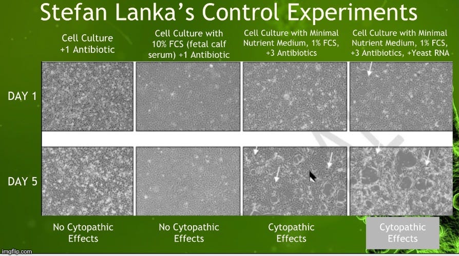
Thus, this cytopathogenic effect that was supposed to be specific to “viruses” and was the sign that a “virus” was present within the culture, could be observed without any “virus” being added to the culture whatsoever. In other words, it was the methods of the experiment itself that created this effect, not the presence of any invisible entity. This finding, which was essentially ignored by Enders, was seen in later experiments by various other researchers (Rustigan, Cohen, Von Magnus, Hull). Even Dr. Lanka himself was able to demonstrate that he could create CPE in a lab using no “viral” materials whatsoever. This finding that the cytopathogenic effect is not specific to “viruses” and can be observed without “viral” materials shouldn’t be surprising as it is well known that CPE can be caused by factors other than “viruses,” such as:
Bacteria
Amoeba
Parasites
Antibiotics
Antifungals
Chemical contaminants
Age and cell deterioration
Environmental stress
It shouldn't be too hard to understand why the results from cell culture experiments are scientifically invalid indirect evidence. First, the assumed “viral” particles are never identified and observed prior to the cell culture experiment taking place. This invalidates the cell culture as a scientific experiment because the independent variable (i.e. the cause - assumed “viral” particles) must be identified before experimentation in order to be varied and manipulated during the experiment. The IV cannot be a creation of the experiment. The dependent variable (i.e. the effect - CPE) must be a naturally observed phenomenon. There is absolutely nothing natural about starving and poisoning cells in a Petri dish.
The second problem, as noted in the highlighted section from Chapter 2 of Introduction to Modern Virology, is that there are “viruses” that are claimed to be uncultivatable, meaning that they do not produce the desired CPE that is meant to indicate the presence of the “virus.” These are known as non-cytopathogenic “viruses,” and this concept makes the cell culture hypothesis that CPE is an indicator of “virus” unfalsifiable as a “virus” can be claimed to be within a culture regardless of the presence of CPE. Interestingly, in a 2021 review of cell culturing in Frontiers, it is stated that cell culturing is “only applicable to CPE-inducing viruses, and therefore it cannot be considered a universal method for virus detection.”
A third unscientific aspect is the lack of proper controls, where samples from healthy hosts are treated exactly as those from the experimental group. These types of controls, which would establish whether or not the methods are causing the CPE rather than the assumed “virus,” are never performed, thus further nullifying the indirect results used to claim the presence of a “virus.”
On top of all of this, the cultures themselves are a mixture of numerous foreign elements and chemicals together, such as the unpurified sample from a human, the kidney cells from an African green monkey, the blood from a baby cow, the addition of antibiotics and antifungals, as well as various other nutrients and chemicals. This process is the exact opposite of purifying and isolating a “virus,” something that HIV discoverer Luc Montagnier stated was necessary in order to prove one has a “real virus.” Thus, the cell culture is a scientifically invalid experimental process that in no way, directly or indirectly, proves the existence of any “virus.”
Indirect Evidence #5: Electron Microscope Images
Particles the size of the assumed filterable “viruses” were not able to be visualized until the advent of the electron microscope (EM) by Ernst Ruska, with his mentor Max Knoll, in 1931. Even then, it wasn't until after Enders established the cell culture technique in 1954 that EM began to be widely used to identify the particles claimed to be “viruses” in the cell culture supernatant. Electron microscopy imaging is the only direct method that virologists use in an attempt to identify their invisible pathogens:
"None of the techniques mentioned so far are able to directly visualise virus particles. That’s where electron microscopy comes in, as it can produce images at the nanometre scale. It does this by firing electrons at a sample and seeing how they interact with it. A computer then interprets this information to produce an image."
https://theconversation.com/five-techniques-were-using-to-uncover-the-secrets-of-viruses-144363
However, the EM process has a while host of issues, and the way it is utilized by virologists ensures that it is an indirect method at best. To begin with, to even obtain images in order to search for the assumed “viral” particles within a sample, the unpurified cell culture supernatant must be put through a series of preparation steps. These steps include dehydration, embedding, fixing, and staining processes that the samples go through before being imaged. According to Dr. Harold Hillman, MB, BSc, MRCS, PhD, these steps significantly alter the sample before it can be viewed. In fact, Dr. Hillman believed that many of the structures seen in EM imagery were, in fact, artifacts created by the various preparation processes. He detailed how far the sample goes from being inside a living organism to the point where it becomes an illustration in a book:
‘What Price Intellectual Honesty?’ asks a neurobiologist
“I was so disturbed by the thought that subcellular fractionation might be an unsatisfactory technique that I decided to take a completely different technique and subject it to a smaller analysis. I took electron microscopy, asking the question, ‘How much does a picture taken with this instrument tell one about the structure of the living cell?’ Since the early 1950s, there has been a passion for relating ‘structure’ to ‘function’, that is, the appearance by electron microscopy of a particular identifiable part of a cell with the biochemistry it exhibits.
The light microscope had been used to examine living cells, unfixed tissue and stained sections, for 100 years until the 1940’s. At that time, the electron microscope was introduced. It permits much higher resolution and magnification than the light microscope, but the tissue can not survive the low pressure, the bombardment of electrons and x-radiation in the electron microscope, so it has to be coated with a deposit of salts of osmium lead or tungsten, which is not destroyed by these agents, and can therefore be examined. Cytologists were very anxious to use this more powerful instrument to look at the fine structure of cells.”
“For example, most cytologists know, but readers of elementary textbooks do not, that when one looks at an illustration of an electron micrograph: an animal has been killed; it cools down; its tissue is excised; the tissue is fixed (killed); it is stained with a heavy metal salt; it is dehydrated with increasing concentrations of alcohol; it shrinks; the alcohol is extracted with a fat solvent, propylene oxide; the latter is replaced by an epoxy resin; it hardens in a few days; sections one tenth of a millimetre thick, or less, are cut; they are placed in the electron microscope, nearly all the air of which is pumped out; a beam of electrons at 10,000 volts to 3,000,000 volts is directed at it; some electrons strike a phosphorescent screen; the electron microscopists select the field and the magnification which show the features they wish to demonstrate; the image may be enhanced; photographs are taken; some are selected as evidence. One can immediately see how far the tissue has travelled from life to an illustration in a book.”
“I have shown, to my own satisfaction that (i) at least some popular important biochemical research techniques have never been controlled, (ii) most of the new structures in cells apparent by electron microscopy are artifacts, (iii) there are only nerve cells and naked nuclei in a ground substance in the brain and spinal cord, (iv) there are no synapses, (v) the transmitter hypothesis is doubtful. I have published all the evidence for these statements, although this has not always been easy.”
https://www.big-lies.org/harold-hillman-biology/what-price-intellectual-honesty.htm
It is easy to understand how the sample is heavily altered before the EM beam even hits it in order to produce the image for the electron microscopist to interpret. These alterations are in addition to the numerous foreign additives and chemicals that were mixed together with the sample during the cell culture process. The images that are assumed to be of “viruses” are not of purified and isolated particles taken directly from the fluids. Instead, they are of unpurifed cell culture soup containing many elements, including cellular debris from the breakdown of dying cells. When the microscopists examine the images, they are searching through a sea of similar and identical particles, trying to find the one particle that represents their preconceived idea of what the “virus” may look like. This can take hours, days, weeks, or longer in order to pick out the representative particle from the mess. According to Luc Montagnier, it “was a Roman effort” that took many hours of searching in order to find the particles, which were not even typical of a normal “retrovirus,” that eventually became known as HIV. When searching for the Marburg “virus,” Werner Slenczka stated that searching for an unknown pathogenic agent in EM can be fatiguing and frustrating as many contaminants can be found within the sample. His team spent more than a day looking for the filamentous particle claimed to be the “virus.” On the second day, the lead technician stepped out for lunch, and when he came back, he was informed that they had found a particle that did not look like any others observed before, and this became their “virus.” When June Almeida found the original “coronavirus” particle in 1967, she searched through unpurified cultures and identified particles that she had seen in murine and chicken samples previously. Her paper was initially rejected by peer reviewers who claimed that the images she produced were just “bad pictures of influenza virus particles.” Thus, it is clear that the particles decided as the “virus” in EM images are nothing but selections based upon the interpretation of the person viewing the images. There is no proof that the chosen particles from the unpurified mess are, in fact, the assumed “virus.”
More damning for EM, the particles claimed to be “viruses” are said to “mimic” normal cellular constituents. Subcellular structures such as multivesicular bodies, clathrin-coated vesicles, secretory vesicles, golgi apparatus, etc. have all been identified as “viruses” by investigators. In fact, many of these normal cellular constituents were recently claimed to be “SARS-COV-2” in various papers. Even worse, the particles claimed to be “SARS-COV-2” were found in EM images of samples that were positive and negative for "SARS-COV-2,” as well as in numerous "SARS-COV-2" negative samples taken from before the "Covid" era. Putting aside the various preparation issues, unless the images of the “virus” come from samples that contain nothing but purified and isolated particles, there is no logical way to conclude that the particles, picked out via what can only be described as the point-and-declare method, are the assumed “virus” over normal cellular components or cellular debris. It is all up to the eye of the beholder. Thus, EM remains, at best, an indirect method of identification of random unpurified particles interpreted as a “virus” by the electron microscopists.
Indirect Evidence #6: “Viral” Genomes & Nucleic Acids
“Detection of viral nucleic acid is not equivalent to isolating a virus.”
-Virologist Charles Calisher
Imagine that someone came up to you with a printout of a genome and told you that it was the embodiment of Bigfoot and that this computer readout was proof that the mythological creature existed. You ask them how they obtained the genome, and they tell you that it was assembled from a mixture of hair, blood, saliva, and feces that were found within the wilderness. You ask them how they know for sure that this collection of unrelated samples came from Bigfoot rather than from an assortment of species, to which they reply that the genome assembled from this mixture has never been seen before. You point out that this would be indirect evidence at best, and that in order to truly know for sure that the genome belonged to Bigfoot, the creature would need to be present in order to obtain the samples from. However, they insist that the long list of A,C,T,G's that you were presented with is evidence enough. This is Bigfoot.
Sadly, this is the circular reasoning employed by many who assert that genomes are evidence of a “virus.” They argue that it does not matter that the actual “virus” has never been purified and isolated directly from the fluids. They claim that a “virus” can be assembled from unpurified fluids, such as bronchoalveolar lavage fluid (BALF) as was done with “SARS-COV-2,” containing many sources of genetic material including host, bacteria, fungus, and other known or unknown elements. They believe that the “virus” can be determined by a process of elimination by comparison to known sequences in a genomic database. They state that any matches to known “viral” sequences demonstrates that they have the genome of a “virus.”
However, this is where the first of many problems arise. No “viral” genome has ever come from purified and isolated particles assumed to be “viruses.” These genomes are always the result of either sequencing from unpurified cell culture creations from a lab or the unpurified samples from a host or the environment. Thus, the entire database made up of known “viral” sequences have never come directly from just the “viral” particles themselves. They are an amalgamation of RNA from many potential sources assembled into a theoretical genome claimed to belong to a fictional “virus.” Just as no one should take the genome of Bigfoot seriously until it is demonstrated that such a creature exists, no one should accept a “viral” genome until the existence of the “virus” has been established and validated scientifically first.
Unfortunately, we currently find ourselves in the age of molecular virology, where the old pseudoscientific indirect methods used to discover “viruses” are being pushed aside for newer pseudoscientific indirect methods claimed to discover “viruses.” We were warned about this looming threat of molecular virology back in a 2001 Science interview with virologist Charles Calisher:
“Calisher has been worrying for years about the wholesale takeover by modern lab toys, fearing that the genetic code they spit out sheds much less light on a class’s workings than “classic” methods. Many senior scientists (some quite a bit younger) share his views: The 14 signatories include some of the most illustrious names in U.S. virology. Together, they have many decades of experience chasing exotic classes across the globe.”
“Nowadays, scientists can detect a virus simply by searching for and amplifjhg snippels of its DNA in human or animal samples. Indeed, they have identified and described quite a few new viruses without ever isolating them.“
Although all that is terrific, says Calisher, a string of DNA letters in a data bank tells little or nothing about how a virus multiplies, which animals carry it, how it makes people sick, or whether antibodies to other viruses might protect against it. Just studying sequences, Calisher says, is “like trying to say whether somebody has bad breath by looking at his fingerprints.“
According to Calisher, studying the A,C,T,G's in a database do nothing to determine if one has a “virus.” These sequences do not show the morphological characteristics of a “virus” nor can they demonstrate that a “virus” is pathogenic. Calisher, along with 13 other virologists of the “old guard,” went even further in their paper that the Science interview was based upon, stating that the “detection of viral nucleic acid is not equivalent to isolating a virus” and that “without an isolate, the pathogenic potential, association with human infections and illnesses, and cross-protectivity are difficult to assess.” This concern on relying on genomic data to tell us anything scientifically was expressed as well by Edward R. Dougherty, the Scientific Director of the Center for Bioinformatics and Genomic Systems Engineering. In a 2008 paper, Dougherty warned that there is an epistemological crisis in genomics, and the major issue is what constitutes scientific knowledge in genomics. He argued that “the rules of the scientific game are not being followed,” and that genomics was amassing a wide collection of data, “but the accumulation of data does not constitute science, nor does the a postiori rational analysis of data.” Dougherty stated that “contemporary genomic research often fails to satisfy the basic requirements” of experimental methods and the scientific epistemology, “thereby failing to produce valid scientific knowledge.” Thus, it is clear that genomic data alone tells us little to nothing about a “virus” nor does the analysis of this data constitute science.
Another issue for “viral” genomes is that, similar to the preparation process utilized by electron microscopy which most assuredly alters the sample in many ways, the various steps utilized to create a “viral” genome does the same. While it is too much to detail here, the processes includes extracting the RNA from the unpurified source through physical and chemical means, shearing and fragmenting the RNA through metal ions or RNAse III, and then converting the RNA into cDNA using the contamination-prone PCR. After this, a library is constructed and analyzed via computer algorithms in order to create the theoretical genome. I detailed this long process here.
This reliance on computer-generated genomes that are indirectly standing in for an entity never shown to exist in a purified and isolated state in order to be sequenced from, has led to the use of fragments of this fraudulent assembly of RNA of unknown provenance to be used as a means of detection via PCR. This has led to disastrous outcomes were the vast majority of the people being tested for and diagnosed with the computer “virus” are entirely free of disease. People have been needlessly locked down, quarantined, and treated for a “virus” that only existed in silico, i.e. in the computer. There is no scientific evidence that links the A,C,T,G's in the computer database to the unpurifued particles chosen in the EM images.
This is just a tip of the iceberg in regard to the many problems with relying on genomics. For a very thorough and in-depth look into these issues, I highly recommend the article DNA Discovery, Extraction and Structure. A Critical Review by Tam. She provides an excellent analysis on why the very foundations of DNA are worthy of being questioned. Whether one believes genomes to be accurate or not, one thing is for certain. Just as in the case of Bigfoot, you can't sequence something that doesn't exist.
The Direct Approach
Hopefully, it is clear now that all virology has to offer in order to claim “viruses” exist are presents under the tree, cookie crumbs with half-drank milk, faint footprints, and questionable security camera footage. What they do not have is Santa Claus, i.e. the “virus” itself. Thus, virologists have established numerous ways to create evidence that implies the presence of an invisible entity so that those who do not question the evidence critically and logically will easily accept that what they are being sold as the truth. It has worked for virology so far, so there is no need for virologists to try and provide direct scientific evidence supporting their claims when the indirect tricks will do.
However, if virologists were honestly interested in proving that their fictional pathogens existed, they would try and establish direct proof of the existence of this entity first and foremost. While they will claim that this is impossible, it should be fairly simple to do so. The first, and most important step, is showing that the particles believed to be “viruses” actually reside within the fluids of a sick human and/or animal where they are supposed to be found in abundance without the use of cell culturing. Virologists have many purification procedures that can be utilized, including ultracentrifugatiion, filtration, precipitation, etc. that can be combined together in order to obtain the assumed “viral” particles away from everything else within the sample. This separation can be confirmed by utilizing EM to show that only isolated particles of the same size and morphology remain after the purification procedures are performed.
Once the virologists know that their sample contains only the particles that they believe to be their “virus,” they can follow the steps of the scientific method and subject a healthy host to these particles via a natural route of exposure. This does not mean injecting the sample into the brains, stomach, veins, testicles, etc. This does not involve scarifying the skin, eyes, mucous membranes, etc. in some way and rubbing the sample into the wound. This means aerosolizing the sample into a mist to be breathed in by the healthy host. Then, the researchers must observe whether or not the same symptoms of disease that they expect to see develop. Obviously, proper controls must be performed throughout the experimentation in order to demonstrate that it truly is these specific particles that are pathogenic. If symptoms of disease do develop, the same purification steps must be performed in order to see if the exact same particles are obtained from the newly sickened host. These particles must then be used in an attempt to sicken another host. This process must be repeated with a large enough sample size in order to ensure accuracy. Once it has been confirmed that the assumed “viral” particles are indeed pathogenic, they can be biochemically characterized and studied further.
If virologists can not purify and isolate the particles directly from the fluids of a sick human or animal, then it's game over. If they do manage success with the first step, but they can not show that these particles are pathogenic naturally via the scientific method, it is also game over. It truly is that simple, and yet, in over 100 years of virology, this process has never once been performed. What we have been given is indirect evidence derived from pseudoscientific methods that, if true, may be used to infer the truth of the matter at hand. What we do not have is direct proof derived from the scientific method that settles the matter that such a thing as a pathogenic “virus” exists. Thus, we must ask ourselves if we are willing to convict the non-existent defendant based off of the indirect pseudoscientific evidence that we have been given, or should we demand that virologists provide the direct scientific evidence that is required in order to prove that the defendant actually exists in order to have committed the crime.
provided an exhaustive list of the various patents and trademarks related to the geoengineering program. got the CDC to admit that there is zero science showing that tick bites lead to disease. created a rather hilarious video for Sense Strand, Bobby Kennedy, Del Bigtree and all the other Virus Pushers Against Clotshots, and members of the Ivermectin Insecticide Cult. examined the media's reporting on Robert Kennedy Jr. and how they have avoided addressing his biggest weakness.











This is a brilliant take-down of viroLIEgy that simultaneously attacks it on all 6 pillars. The useful idiots of virology certainly deserve blame but the real culprits are the PTB who funded it and determine the "consensus".
I am a detective and your investigative work is astounding. I consider you a fellow detective.