“You go through narrow-pored clay filters, which hold back all bacteria, easily pass through and you have they are not yet visible with the best microscopes, including the ultramicroscope can do. We must infer their existence because they represent various human, animal and plant diseases. It's a very special strange fact that we are dealing with these microorganisms that are completely invisible to us can operate in exactly the same way as with pure cultures of bacteria.”
-Robert Koch
The above quote was taken from one of German bacteriologist Robert Koch's final speeches, the inaugural address at the Academy of Sciences on July 1, 1909. He passed away almost a year later on May 27th, 1910. At the time, Koch acknowledged his belief that there were entities that were invisible even under the best microscopes. As they were invisible and represented certain diseases, their existence had to be inferred from evidence that was similar to that seen in the studies on bacteria. In other words, if a bacterium was sought after and failed to be identified as the causative agent of a disease, it was acceptable to blame an unseen culprit. The diseases that could not be linked to bacteria and required the invisible scapegoat to keep the germ theory alive included measles, scarlet fever, smallpox, rabies, influenza, yellow fever and cattle plague. According to Field's Virology textbook, the concept of the invisible “virus” was born once the researchers realized that they were unable to satisfy Koch's Postulates, the criteria considered absolutely necessary to fulfill in order to prove that microbes cause disease:
“These studies formalized some of Jacob Henle's original ideas in what are now termed Koch's postulates for defining whether an organism was indeed the causative agent of a disease. These postulates state that (a) the organism must be regularly found in the lesions of the disease, (b) the organism must be isolated in pure culture, (c) inoculation of such a pure culture of organisms into a host should initiate the disease, and (d) the organism must be recovered once again from the lesions of the host. By the end of the 19th century, these concepts became the dominant paradigm of medical microbiology. They outlined an experimental method to be used in all situations. It was only when these rules broke down and failed to yield a causative agent that the concept of a virus was born.”
Researchers began to claim that, if they used filters that were small enough to keep known bacteria out, and the resulting fluids after filtration resulted in symptoms of disease in animals, this was evidence that something smaller than a bacteria existed within the fluids that caused the disease. This gave rise to the term “filterable viruses.” The Field's Virology textbook goes on to explain that, once this idea of “filterable viruses” was accepted, a procedure was created in order to find them. This is the technique known as the cell culture that was established by John Franklin Enders in 1954, nearly 60 years after the idea of the “filterable virus” was conjured up. Virologists had to rely on factors such as the size of the pore of the filters, whether there was a reaction to chemical agents (alchohol and ether), and whether or not they observed cytopathogenic effects (CPE) in the cell culture as indirect evidence (i.e. evidence that does not prove a fact but can be used to infer that the fact exists) in order to claim that the invisible entities were within the fluids. As virologists could not see the entities that they assumed to be present, they had to rely on faith that they were there:
“Once the concept of a filterable virus took hold, this experimental procedure was applied to many diseased tissues. Filterable agents, unable to be seen in a light microscope, that replicate only in living animal tissue were found. There were truly some surprises, such as a virus—yellow fever virus—transmitted by a mosquito vector ( 122), specific visible pathologic inclusion bodies (viruses) in infected tissue (80,116), and even viral agents that can “cause cancer” ( 43,123). Throughout this early time period (1900–1930), a wide variety of viruses were found (see Table 1) and characterized with regard to their size (using the different pore sizes of filters), resistance to chemical or physical agents (e.g., alcohol, ether), and pathogenic effects. Just based on these properties, it became clear that viruses were a very diverse group of agents. Some were even observable in the light microscope (vaccinia in dark-field optics). Some were inactivated by ether, whereas others were not. The range of viral diseases affected every tissue type. Viruses gave rise to chronic or acute disease; they were persistent agents or recurred in a periodic fashion. Viruses might cause cellular destruction or induce cellular proliferation. For the early virologists, unable to see their agents in a light microscope and often confused by this great diversity, there had to be an element of faith in their studies. In 1912, S. B. Wolbach, an American pathologist, remarked, “It is quite possible that when our knowledge of filterable viruses is more complete, our conception of living matter will change considerably, and that we shall cease to attempt to classify the filterable viruses as animal or plant”
The concept of the invisible “virus” was used in order to explain away any evidence that contradicted the idea that microbes were the cause of disease once it was realized that Koch's logic-based requirements could not be satisfied. Virologists were emboldened to bend the rules as Koch himself regularly did so as well. He knew that it was often impossible to induce disease in animals in order to claim a microbe as the causative agent, as evidenced by his troubles with cholera. Unfortunately, instead of realizing that his Postulates worked as designed by disproving microbes as causative agents of disease, Koch allowed for logic to be bent in order to keep the germ theory alive. This was admitted in Alfred Grafe's “A History of Experimental Virology:”
“Since Koch knew, after his 1884 experience with cholera, that it was often impossible to induce a disease experimentally in animals and yet not harbour the slightest doubt about the germ theory, he augmented these guidelines. His thoughts on the implications of the regular and exclusive occurrence of bacteria in infectious diseases without a possible animal experimental trial was reflected in his own words:
"Our contention is likely justified, even at this point, that if only the first two requirements for proof are fulfilled ... the causal relationship of parasite to the disease is validly established."
Nevertheless, it is essential to explain how it was possible to arrive at Koch's postulates in a manner which contradicted Koch himself.”
As admitted by Grafe, Koch completely contradicted himself by allowing for a microbe to be claimed as a causative agent even if the disease was not recreated experimentally. Previously, Koch claimed that this was the only possibility of providing direct proof of causality:
“The only possibility of providing a direct proof that comma bacilli cause cholera is by animal experiments. One should show that cholera can be generated experimentally by comma bacilli.”
-Robert Koch
Koch, R. (1987f). Lecture cholera question [1884]. In Essays of Robert Koch. Praeger.
Koch also eventually allowed for the microbe to be identified as the causative agent in cases where the disease did not occur (i.e. in the healthy). Thus, depending on the situation, Koch abandoned and contradicted his own logical postulates in order to fit evidence to the germ theory of disease so that it could be kept afloat in the face of contradictory evidence and the inability to fulfill his criteria. The creation of the invisible “virus” was the latest effort to try and plug the holes in the sinking germ theory ship.
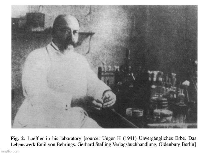
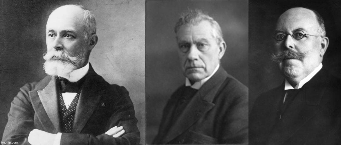
The creation of this concept of the “filterable virus” is primarily credited to three different researchers: Dmitri Ivanovski, Martinus Beijerinck, and Friedrich Loeffler. The former two were involved in the “discovery” of the first “virus” ever identified known as the tobacco mosaic “virus” (TMV) that afflicts certain plants, while the latter was involved in the “discovery” of the first vertebrate “virus” with the foot-and-mouth disease “virus” (FMD). Let's explore the work of these men that led to the formation of this “filterable virus” concept, and see whether or not this idea emerged from scientific methods.
Tobacco Mosaic “Virus” (TMV)
Tobacco mosaic “virus” is regarded as the first “virus” ever to be discovered, officially ushering in the era of virology. This is a disease that is supposed to result in spotting discoloration of the leaves of the plant. However, the symptoms experienced (mottling, yellowing, leaf curling, stunted growth, and necrosis) by the plant are said to be very dependent on the host plant, the age of the infected plant, the environmental conditions, and even the genetic background of the plant. These influences on the disease process were relegated to co-factors as the search for a specific microbe was pursued. Due to the inability to cultivate a bacterial agent that could be associated with the disease, the idea floated about that there was an invisible entity, originally regarded as a poison rather than actual particles, that passed through filters small enough to keep out bacteria.
With TMV, the two men often credited with the discovery of the first “virus” are the aforementioned Dmitri Ivanovski and Martinus Beijerinck. In 1892, Ivanovski attempted to discover the microbial cause of the disease by crushing the leaves of diseased plants and passing the resulting leaf sludge through various filters. He claimed that the filtered juices, when inoculated onto healthy plants, produced the same disease as seen in nature. This usually involved scraping or injecting the plant and/or leaves, thus damaging them, putting the filtered juice on the leaves, and then monitoring the leaves to see if the spotted disease occurred. Ivanovski's 1892 paper “Considering the Tobacco Mosaic Disease in Plants” is considered the first report on the filterability of “viruses.” However, there are no details on his experimental methods within the paper, and all that is provided are his own claims that passing the crushed plant leaves through a Chamberland filter, said to hold back bacteria and fungi, still produced disease:
“that the sap of leaves attacked by the mosaic disease retains its infectious qualities even after filtration through Chamberland filter candles.”
According to Alfred Grafe's “A History of Experimental Virology,” Ivanovski concluded that the agent must be either a specific bacterium or a specific toxin, although he could find neither. Even upon further experimentation by Ivanovski in 1903, and after assessing the results of his experiments on the tobacco mosaic disease agent, Grafe concluded:
“it is not possible to find evidence on the nature of the causative agent. Ivanovski did not offer proof in his experiments or in his illustrations that the causal agent was a bacterium. Furthermore, there is no indication of his having suspected a new type of causative agent.”
Thus, it is easy to see that Ivanovski could not identify any agent—bacterial, fungal, or “viral”—and relied on lab-created experimental effects, such as filterability, in order to claim that an invisible “infectious agent” was present. He remained convinced that, despite being unable to cultivate any bacterium along with repeated failures to produce evidence, the causal agent was an unculturable bacterium that was too small to be retained on the Chamberland filters or to be detected by light microscopy. Regarding the idea that the causative agent could be something other than bacterial, Ivanovski claimed that he:
“succeeded in evoking the disease by inoculation of a bacterial culture, which strengthened my hope that the entire problem will be solved without such a bold hypothesis.”
Ivanovsky, D. 1899 Ueber die Mosaikkrankheit der Tabaksp£anze. Centbl. Bakteriol. 5, 250^254
This was Ivanovski's response to the idea proposed by Martinus Beijerinck a few years later in 1898 that the “filterable agent” of the tobacco mosaic disease was not bacterial, but rather something like what we now think of as a “virus,” which he called the Contagium vivum fluidum, i.e. contagious living fluid. In his work, Beijerinck repeated a similar process to Ivanovski and attempted to filter the juices of diseased plants and then inoculcate the healthy plants with injections of the filtered fluids. The leafs that were “infected” occurred directly above the “wound” created by the syringe, which is not a natural route of exposure or “infection.” In order to speed up the disease, all one had to do was create a deeper wound and insert diseased material. Interestingly, Beijerinck discussed how formalin used to sterilize the syringe was extremely toxic to the tobacco plant and that one had to ensure that no traces of formalin remained in the syringe when used for experiments. He even mentioned that the disease from the artificial inoculation was different from the disease that is seen naturally in plants:
Concerning a Contagium vivum fluidum as cause of the spot disease of tobacco leaves
“The quantity of candle filtrate necessary for infection is extremely small. A small drop put into the right place in the plant with a Pravaz syringe can infect numerous leaves and branches. If these diseased parts are extracted, an infinite number of healthy plants may be inoculated and infected from this sap, from which we draw the conclusion that the contagium, although fluid, reproduces itself in the living plant.”
“Often (perhaps always) the leaf that first becomes diseased is situated directly above the wound left by the infecting needle. If the place of infection was closely circumscribed, for example to a single shallow puncture of the needle with the Pravaz syringe, the second diseased leaf, in a s leaf position, may be exactly the ninth above the first one to become diseased.”
“If one wishes to convince oneself in the shortest possible time of the virulence of the contagium it is best to deeply wound with a knife the youngest part of the stem below the terminal bud, which still may be easily treated without injury, and to place into the wound a piece of fresh, diseased tissue. The newly formed leaves will then plainly show the first traces of the disease after ten to twelve days; after three weeks the disease symptom is clearly distinguishable, even to the layman.”
“In any case, one must be sure that the last traces of Formalin have completely evaporated from the syringe before using it again, for it has become apparent that Formalin is very poisonous for the tissues of the tobacco plant, much more so than to the virus itself.”
“Although most of the dead tissue spots develop in the manner described near or in the dark-green fields near the veins, the origin of some of them remains uncertain; apparently, they also may develop in the yellow spots. The symptoms in the tobacco fields are usually not of as great an intensity as in artificial infection, especially the blistery outgrowth of the dark-green parts on the leaf blade is entirely lacking. In contrast to this, the necrosis and drying of leaf spots were not observed in some of the greenhouse plants.
With artificial injection of fresh extracted sap, or with inoculation with diseased tissue the disease may reach a higher stage of intensity than I have as yet observed under natural conditions. I mean the abnormal tissues of the newly formed leaves (Plate I b, c, d, Plate II, fig. 4 and 5). This is no doubt connected with the quantity of infectious material used for the experiment. Therefore, it is much easier to produce leaf monstrostities with fresh extracted juice than with the Bougie filtrate, since, as has been remarked earlier, more of the latter must be injected m order to obtain the same effect, which certainly is remarkable for a contagium that increases through growth.”
Being unable to produce the disease exactly as seen in nature along with the unnatural route of “infection” by wounding plants and injecting them from a syringe should be enough to disqualify Beijerinck's experimental results and conclusions. However, like the virology papers that came afterwards, no proper controls were ever performed by Beijerinck with fluids from healthy plants treated and inoculated in the same manner, thus further disqualifying his findings. According to Grafe, Beijerinck's ideas about the causative agent, which he regarded as a liquid toxin rather than infectious particles, were disregarded, rejected, and no longer discussed at the time:
“Beijerinck's concepts were equally disregarded! As early as 1898, he had assumed in his reflections on colloid chemistry that "virus" might be a living, liquid contagium, and commented that it was probably taken up by a living cell, in which it then reproduced. Although protein-like crystals, i.e. organized biological material, had been known since Hartig's description ofthem in 1856, a liquid contagium hardly fitted into the concept of an infectious pathogen at that time. In short, Beijerinck's idea met with rejection and his model of multiplication was discussed no further.”
Interestingly, when attempting to determine a “founder of virology,” Grafe mentioned that even though Beijerinck's ideas were close to the concept of the “virus” as it is known today, he made no attempt, theoretical or experimental, to prove or even to defend his hypothesis that the infectious agent was a contagious living fluid. In other words, all Beijerinck had was an unproven hypothesis, i.e. an assumption, which essentially disqualifies him. Grafe equally questioned Ivanovski as “the founder of virology” as, in order to even consider him, all of Ivanovski's published findings would need to be disregarded. Grafe stated that Ivanovski did not follow the established rules of etiological experiments and arrived at false conclusions. Thus, the two men that are most often credited as having discovered the first “virus,” are completely discredited as neither were able to identify any causative agent:
The literature often cites someone as the "founder" of virology. Since it is customary - for non-scientific reasons - to associate the word "founding" with one particular person, in the case of virology such a contention can only be defended superficially. When Beijerinck is mentioned, the scales tip in his favour because his ideas at the close of the 19th century sound to us so modern. Speaking against him is the fact that he made no attempt, theoretical or experimental, to prove or even to defend his hypothesis of a Contagium vivum fluidum and its intracellular reproduction. To declare Ivanovski "father of virology" can only be credible if we disregard his publications. He did not observe the established rules for etiological experiments, and consequently arrived at false conclusions. In his opinion, the pathogen for TMD was a bacteria which could be photographed. On the other hand, no one can deny that he was the first to filter the causative agent of a plant disease, not discounting Mayer's use of filter paper in 1880.”
Foot-and-Mouth Disease (FMD)
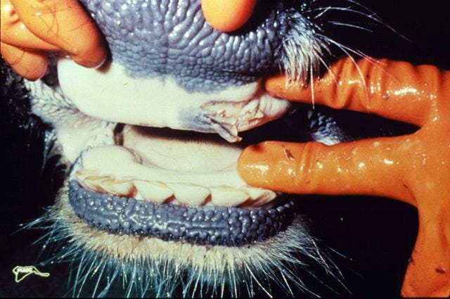
Foot-and-mouth disease (FMD) is a set of symptoms said to first be identified as a specific disease in the late 1800s. According to the Cornell College of Veterinary Medicine, FMD is characterized by fever and blister-like lesions followed by erosions on the tongue and lips, in the mouth, on the teats, and between the hooves. Most animals recover, but the disease “results in a weakened state, loss of weight, and reduced production of milk and meat.” It is said to afflict mostly animals such as cows, pigs, sheep, goats, deer, and others with divided hooves, but it has been claimed to affect humans as well. As this disease was affecting livestock and causing economic loss, there was a major push by the Prussian government to find a vaccine against it. This ultimately led to the appointment of Friedrich Loeffler, a pupil of Robert Koch, to head up a Commission established n 1897 seeking to create a vaccine against the disease. He was under considerable political and economic pressure to create a vaccine as quickly as possible, even without identifying any specific cause.
The results from Loeffler's Commission were reported in four separate documents between April 17, 1897 and August 12, 1898. In the last of Loeffler's four reports, he called attention to the fact that there were several bacterial and other amoeba-like microbes claimed by various researchers to be the causative agents of FMD. He spent a considerable amount of time at the beginning of his report discussing the work of Drs. Siegel and Bussenius, who had claimed to have discovered a specific bacterium in man and animals that had died of the foot-and-mouth disease. The doctors claimed to have even recreated the foot-and-mouth disease experimentally in animals using pure cultures of their bacterium. However, when Loeffler's Commission attempted to grow the same bacterium from the blood of sickened animals, the results were considered negative, even though certain bacteria were grown. These were claimed to be either micrococci or pseudo-diphtheria bacilli that were said to be impurities due to contamination.
The Commission also attempted to recreate Drs. Siegels and Bussenius’ animal experiments, even though they had the excuse at the ready for any experimental results that may have confirmed the findings. They stated that experiments of this sort “might easily lead to a false conclusion, since the disease might be conveyed to the inoculated animals by the attendants, or even by the members of the Commission themselves.” In other words, any positive results may not be due to a bacterium but by accidental spread of the “actual causative agent” by those involved. Attempts were made to “infect” calves by feeding them 50 ccm. of two-day old bouillon or by inoculation from scarification (involves scratching, etching, burning/branding, or superficially cutting) on the upper and lower lips. This was said to be the same method used to “infect” animals with foot-and-mouth disease lymph. The sucking calves became ill with high fever and symptoms of intestinal affection, with one killed while the other died “naturally.” The bacilli were found in the blood and spleen of both animals. However, as neither animal had the characteristic lesions on the mouth or hooves, the experiment was considered negative.
Due to concerns by Drs. Siegel and Bussenius that not enough time was given for the lesions to develop, three more calves were experimented on. However, the amount of bouillon that was used with the calves was reduced from 50 ccm to 2 ccm in one calf and 5 ccm in the two remaining calves. This produced similar results and, once again, the experiments were considered negative. Loeffler concluded that Drs. Siegel and Bussenius had an “interesting and remarkable pathogenic organism” that was “capable of setting up severe intestinal disease.” However, it was decided not to be the cause of foot-and-mouth disease. Somehow, these results were also used to disregard the findings of a bacterial cause by numerous other researchers (Nosotti, Klein, Schotteluis, Kurth, Nissen, Starcovici, Furtuna, and Stutzer) as well. Even claims put forward by Piana, Fiorenti, Behla, and Jurgens of small protoplasmic structures with distinct amoeboid movements as causative agents were disregarded in favor of Loeffler's own unidentifiable and invisible agent:
Report of the Commission for the Investigation of Foot-and-Mouth Disease at the Institute for Infectious Diseases, Berlin.
“Nevertheless the Commission considered it necessary to submit to examination a particular species of bacterium which had been found by Drs Siegel and Bussenius in alleged fatal cases of foot-and-mouth disease in the human subject, and also in cases of the same disease in animals, and this appeared to be the more necessary because these authors had alleged that they had been able to produce typical foot-and-mouth disease in calves and pigs with pure culture of their bacillus.”
“Inasmuch as, according to the views of its discoverers, the bacillus is mainly found in the blood of recently attacked animals, although frequently only in very small numbers, special attention was always directed to examination of the blood. In five cases blood was taken from the jugular vein of recently attacked animals by means of a sterile trocar, and collected in sterile Erlenmeyer flasks; blood was also taken from the heart of two calves which had been killed at the height of the disease immediately after development of vesicles. Large quantities of the blood were used to inoculate bouillon, nutrient agar, and nutrient bouillon, the flasks being then placed in the incubator. In the great majority of cases the flasks thus inoculated remained permanently sterile, but in a few of them micrococci, and in some others bacilli, developed. Most of the latter belonged to the group of pseudo-diphtheria bacilli, and had not the most remote resemblance to the bacillus of Siegel and Bussenius. They were obviously accidental impurities which had been obtained from the skin of the animals in taking the blood.
These negative results of experiment are in contradiction with the positive assertion of the authors named, that they had succeeded in producing the typical disease with their bacillus; nevertheless the Commission considered it advisable to afford them an opportunity to demonstrate the experimental production of the disease with the bacillus. The Commission were mindful of the fact that experiments of this sort might easily lead to a false conclusion, since the disease might be conveyed to the inoculated animals by the attendants, or even by the members of the Commission themselves, who were almost daily brought mto contact with animals suffering from foot-and-mouth disease.”
“Inasmuch as it was possible that the bacillus of Seigel and Bussenius might have lost some of its virulence from long cultivation in nutrient gelatine, at the desire of these two gentlemen an attempt was made to infect the two sucking calves by pouring about 50 ccm. of a two-days-old bouillon culture of the bacillus into the mouth of each animal. At the same time, however, two yearlings were inoculated by scarification on the upper and lower lips in the same manner as one proceeds in infecting animals with foot-and-mouth disease lymph. In the case of the yearlings the material employed was a fresh culture of agar, and a large quantity of the culture was also rubbed on the mouth of these two animals, so that an infection of the intestine was also made possible.
On the following day the sucking calves were already ill, with high fever and symptoms of intestinal affection. One of them was killed while very ill on the third day, and the other died in the course of the following night. In both of these animals the bacilli were found in the blood and spleen, and especially in the much swollen mesenteric glands, as well as in contents of the intestines. Their presence in these positions was demonstrable by microscopic and cultural examination. Neither of the animals had lesions in the mouth or on the feet, such as are characteristic of the disease in question. On the contrary, they were affected with a severe enteritis.
With a two-days-old bouillon culture started from the heart blood of the above calf in which death resulted naturally, three new animals were infected at the instigation of Drs Seigel and Bussenius, in order to induce a less acute form of the disease, and thus to give time for the production of the characteristic lesions. One of these was a sucking calf and it received 2 ccm. into the mouth. The second animal was a three-months-old calf, and the third a yearling, and each of these received 5 ccm. into the mouth. On the following day the sucking calf was already the subject of high fever and profuse diarrhrea, and it died on the fourth day. The post-mortem examination showed practically the same conditions as in the above-mentioned calves.”
“The three-months-old calf became ill on the second day, being also attacked with high fever and profuse diarrhrea. Subsequently, however, it recovered, though its temperature remained for a long time over 41° C. Both the inoculated yearlings, and also the one infected by feeding, sickened on the fourth day, with rather high fever and profuse diarrhrea. The fever lasted from two to four days, and as it declined the diarrhrea also abated. During fourteen days' observatIon none of the animals showed any symptoms of foot-and-mouth disease.
From these experiments it follows that the bacillus of Seigel and Bussenius, although an interesting and remarkable pathogenic organism, capable of setting up severe intestinal disease, is not the cause of foot-and-mouth disease.
From the results of the investigations above described it may also be concluded that the bacteria found by other observers-Nosotti, Klein, Schotteluis, Kurth, Nissen, Starcovici, Furtuna, and Stutzer, in cases of foot-and-mouth disease, did not represent the causal agent of the infection.
There still remained for investigation the claims of certain observers who have described, not bacteria, but small protoplasmic structures with distinct amoeboid movements, as the cause of the disease. Claims of this sort have been put forward by Piana, Fiorenti, Behla, and Jurgens.”
According to Loeffler, the only animals that he could “infect” with any certainty with his “agent” were calves. Experiments with twenty-two pigs only resulted in eight “infections.” He admitted that there were no successes in eight attempts with sheep, and only one out of eight goats became “infected.” He also tried, via inoculation into the mucous membrane of the mouth or the limbs, intraperitoneal injection, or through the feeding with fresh materials, to “infect” 30 rabbits, 14 guinea-pigs, 3 dogs, 4 cats, 5 rats, 10 house mice, 10 field mice, 6 hens, and 6 pigeons, but all of these attempts failed.
Loeffler stated that the way to “infect” animals was with rubbing the mouths with materials moistened in the fluids of deceased animals. However, he admitted that this did not always result in “infections.” In one instance, only 9 of 17 animals were said to be “infected” in this manner after three days of attempts. The disease was said to be successfully experimentally transmitted to two yearlings and 13 calves by rubbing a drop of lymph on the slightly scarified mucous membrane of the upper and lower lips. In other words, they roughed up the mucous membranes in some way (cutting, sanding, burning, etc.) and then rubbed lymph into the wound. If blisters formed after abrasive scarification and the rubbing of unclean materials into the wounds, it was considered a success. No attention is paid to the very acts of scarification and rubbing being the cause of the blisters rather than any invisible entity. In the great majority of the cases, the disease only attacked the areas wounded around the mouth and not the feet of the calves.
Transmission of the Disease to Different Species of Animals.-The only animals that we have been able to infect experimentally with certainty are cattle (2) or calves (13). Out of twenty-two attempts to infect pigs only eight were successful. There was no success in eight attempts with sheep, and only one out of the same number with goats. Attempts were also made to infect 30 rabbits, 14 guinea-pigs, 3 dogs, 4 cats, 5 rats, 10 house mice, 10 field mice, 6 hens, and 6 pigeons, but the attempts failed, whether they took the form of inoculation into the mucous membrane of the mouth or into the limbs, intraperitoneal injection, or feeding with fresh materials.
Mode of Infection.-According to experience, in adult cattle and calves infection follows when materials moistened with fresh slaver from diseased animals, such as towels, sponges, or wisps of straw, are used to rub the mouth; but even in this way infection does not always follow with certainty. For example, in one instance only nine out of seventeen animals which it was sought to infect in this way on each of three successive days in the same building contracted the disease. The disease was experimentally transmitted to two yearlings and 13 calves by rubbing a drop of lymph on the slightly scarified mucous membrane of the upper and lower lips. The vesicles developed not only at the places scarified but also in their neighbourhood, on the palate and tongue. In the case of the yearlings vesicles developed on the feet, but in the great majority of cases the disease did not attack the feet in the calves. It was observed that the vesicles formed on the feet one or two days after the disease had shown itself in the mouth. On the second or third day after experimental infection the temperature rose 1° or 1 1/2°, and in the course of the next three days it fell to the normal. The formation of vesicles set in from the first to the third day, generally on the second day, after inoculation. In the case of ten oxen inoculated in the skin of the back or on the posterior aspect of the thigh, no local lesion was produced, and up to the fifth day after inoculation none of the animals appeared to have contracted the disease. Unfortunately this experiment was vitiated by the fact that three days after the inoculation in the skin the owner had also attempted to infect the animals, so as to get rid of the disease as quickly as possible. After that the animals developed the symptoms of foot-and-mouth disease, but it was impossible to determine whether this was the result of the first or the second attempt to infect them.”
The material said to be “infectious” were materials taken from the vesicles. Attempts to “infect” with the serum of the blood from diseased animals failed to produce disease. The most certain method of “infection” was considered the introduction of the invisible “virus” hiding within the contents of vesicles into the blood stream. Loeffler also claimed success by injecting the “virus” into the peritoneal cavity and into the muscles, as well as rubbing the materials into the mucous membrane of the mouth after wounding by pricking. Inoculations into and under the skin proved uncertain:
“Infective Material.-The contents of freshly developed vesicles have been found to be the most certainly effective material for infection. Serum of blood taken from the jugular vein of diseased animals during the period of vesicle formation, and subcutaneously injected in quantities of from 10 to 14 ccm., did not produce the disease in three calves. Experiments regarding the infectivity of the urine and feces of diseased animals have not yet been carried out.”
Method of Infecion.-Through numerous comparative experiments it has been ascertained that the most certain method of infection is the introduction into the blood stream of the virus found in the contents of vesicles. Success has also attended the introduction of the virus into the peritoneal cavity, injection into the muscles, and rubbing of it into the mucous membrane of the mouth after wounding by pricking. On the contrary, inoculations into and under the skin have proved uncertain. Both of these appear to be effective only when at the same time the virus is introduced into a blood vessel. From the moment that the temperature begins to rise till the development of the local symptoms of the disease, the virus circulates in the blood stream, but it disappears from the blood after the development of the local lesions. The disease could be conveyed to healthy animals with from 50 to 100 ccm. of blood taken from the jugular vein of an animal twenty to twenty-eight hours after its inoculation.”
In attempts to determine the microbe via injections of filtered and unfiltered lymph into animals, both the filtrate and the control animals showed typical signs of illness. When “fresh lymph fluid” was used, the animals treated with the filtered lymph always became affected in the same way as the control animals treated with unfiltered lymph. This led Loeffler to two possibilities: either the “germ-free” filtered lymph contained an “extraordinarily active poison,” or there was an undiscoverable agent that was so small that it could pass through the pores of a filter that bacteria could not pass through:
“The result of these injections was somewhat surprising. The animals which received the filtrate contracted the disease in the same time as control animals which had received a corresponding quantity of unfiltered lymph, and they all showed typical symptoms of the disease, with high fever and the development of vesicles in the mouth and on the feet. It appeared as if the activity of the lymph had not been affected by the filtration. In order to be quite certain regarding this point the experiments were several times repeated on a large number of calves and pigs. When fresh lymph was used the results were always the same, the animals treated with the filtered lymph always becoming affected in a quite typical way, just like the control animals treated with unfiltered lymph.
How was this striking fact to be explained? Two alternative explanations offered themselves- either the germ-free filtered lymph contained in solution some extraordinarily active poison, or the hitherto undiscoverable agent of the disease was so small that it could pass through the pores of a filter which will certainly keep back the smallest of the known bacteria. If the first explanation is the correct one, the soluble poison must be an amazingly active one.”
According to Grafe, Loeffler's FMD reports resulted in a “pathogen” that was “not identifiable under the microscope” and was one “belonging to a new type of causal agent.” The Commision decided that, in order to develop a vaccine, it was desirable—but not absolutely necessary—to identify the causative agent. Loeffler and his colleagues concluded that the FMD “pathogen” belonged to a group of the smallest organisms, but Grafe pointed out that they did so “without any clear demarcation to the bacteria.” Grafe stated that the criterion of a "filterable virus" was considered significant by Loeffler as “a laboratory index but not for the characterization of a new causative organism.” In other words, Loeffler did not clearly distinguish between bacteria and his “filterable agent,” and only used the term to classify his unidentifiable agent rather than claiming that a new entity existed.
A 1999 paper by H. P. Schmiedebach provides even more details on Loeffler's Commission and their work, highlighting many other reasons for why their results can be disregarded. The primary goal of the Comission was to find a vaccine. The identification of the “causative agent” was considered a side effect from these efforts. However, as it was not possible to actually visualize the “virus,” Koch's Postulates could not be completely employed. Schmiedebach noted that Loeffler's work de-emphasised the theoretical and the scientific requirements while the political, practical and public context ultimately determined the experimental strategy as well as the focal points of his research. Therefore, Loeffler could not present scientifically derived evidence for the existence of his unidentifiable agent.
Loeffler excused his inability to directly identify his agent by guessing that it must be smaller than 0.1 um, thus even the best microscopes of the time would not be able to visualize it. There were no means by way of microscopy or any other methods that the Commission could utilize to identify the invisible microbe. Oddly enough, in 1903, Loeffler declared Koch's Postulates as the decisive goal to be met in order to prove a microorganism causes disease. Ironically, without being able to visualize the entity nor grow it in pure culture, Loeffler failed to satisfy this “decisive attainment” with regard to the identification of the FMD microbe:
The Prussian State and microbiological research - Friedrich Loeffler and his approach to the "invisible" virus
“The research programme concerning FMD, which was initiated by political authorities, was primarily aimed at the development of an effective vaccine but led, as a side-effect, to the virus itself. Nonetheless, it was not possible to find a way to visualize the supposed virus and thus Koch's postulates could not be completely employed. Loeffler emphasized the practical side of his research activities, which to some extent de-emphasised the theoretical and scientific requirements. The political, practical and public context determined the experimental strategy and the focal points of his research.”
“These calculations, based on a mechanical view without consideration of possible metabolic activities, brought him to the conviction that there existed a germ of very small size that could pass through common filters. Nonetheless, he could not present scientifically derived evidence for the existence of that small germ. Loeffler was fully aware of this epistemological dilemma. In his record he added an explanation for the impossibility of seeing this very tiny germ. He referred to correspondence with Professor Abbe in Jena who was an authority of the highest reputation regarding microscopic techniques. Loeffler discussed with him the limitations of microscope performance. If the supposed germ of FMD had a size of only about 0.1 um, even the best immersion techniques of that day could not made this virus visible. According to Loeffler, this would be best explanation for the fruitless attempts to discover the germ by microscopic investigation. Although this was a very pessimistic view, he tried to turn the tables and offered a perspective concerning the possible discovery of a large number of other germs that could not be identified at that time. In connection with the necessity of future studies on that problem, the commission also requested for the grant of new financial support from the government.”
“The commission could not identify the supposed microbe by microscope nor was it possible to make it visible by any other methods. However, there were some scientific requirements to be fulfilled in order to accept a supposed germ as the causative agent for a disease. In 1903 Loeffler himself wrote on the occasion of Koch's 60th birthday about the scientific foundations of the newly emerged discipline of bacteriology and declared the development of "Koch's Postulates" as a decisive attainment. While Koch had mentioned four postulates in 1890, Loeffler referred only to three:
1. "Constant evidence of the concerned organism in all cases of the disease;
2. isolation of the pathogen in a pure culture that had to be cleaned of all corpuscular elements of the sick individual;
3. generation (Wiedererzeugung) of the disease anew by reliable pure cultures."
I will not discuss the differences from Koch's original formulation but we must evaluate whether Loeffler himself undertook any steps to employ these three postulates in the case of FMD and which, if any, he employed. As we have already seen, two very successful methods, microscopy and culture, failed with regard to the identification of the microbe.”
Interestingly, Schmiedebach noted that, in 1907, the Prussian government accused Loeffler of spreading FMD via his experiments, and he was shut down for two years. Loeffler did not publish any papers describing the nature of his “virus” and he performed no experiments to prove any theories in the field. Loeffler was tied to the bacteriological field as taught to him by his mentor Koch, and he saw no convincing reasons to reject his teachings:
“In 1907, Loeffler was confronted with numerous difficulties. Since 1902, he had been using a farm in Greifswald for his trials. In 1907, the Prussian Minister of Agricultural Affairs accused Loeffler of being responsible for the dissemination of FMD in the region of Greifswald. Loeffler's experiments at this farm were seen as the main cause for the spread of the disease.”
“Following this, and considering the heavy pressure, it is not surprising that Loeffler was not in the contemplative mood required to publish sophisticated articles about the nature of the virus and to perform experiments to prove one or the other theory in this field. In addition, he was adherent to the bacteriological paradigm that had presented many successful discoveries up to that time and he did not see any convincing reason to reject this concept. Considering all the ramifications of his work, his research strategy, despite all the remaining questions and the epistemological uncertainty, becomes more comprehensible.”
https://pubmed.ncbi.nlm.nih.gov/10470267/
According to chapter 3 of Grafe's A History of Virology, Loeffler’s methods were the model for others who investigated “filterable viruses” at the time. Even though the term included the word “virus,” the opinion prevailed for over three decades after it came into use that the “filterable virus” was merely a very small bacteria. Nothing was undertaken experimentally to investigate other hypotheses that differentiated the “filterable viruses” from being anything other than small bacteria.
Thus, it is abundantly clear that, like Ivanovski and Beijerinck before him, Loeffler did not discover any new causative organism. Even though he considered Koch’s logic-based criteria as decisive in proving a microorganism as the cause of disease, Loeffler was unable to satisfy Koch's Postulates for his “filterable virus.” He was unable to culture any microorganism nor could he visualize one. His experimental “infections” resulted in contradictory results depending on the species of animals and the route of “infection.” Reminiscent of Ivanovski and Beijerinck's own methods, no attention was given to the fact that the best results came from unnatural routes of exposure that required wounding the host. With Ivanovski and Beijerinck, this meant cutting the plant while injecting materials into the resulting hole. For Loeffler, this meant scarifying the lips, mouths, and mucous membranes of the animal and rubbing diseased materials into the wounds. These methods do not reflect nature in any shape or form.
Regardless, none of the three researchers credited with identifying the “filterable viruses” for the first time provided valid scientific evidence to support such a claim as to the discovery of a new entity. Dmitri Ivanoski's methods were ill-defined and he did not follow the established rules of etiological experiments, leading to false conclusions. He did not provide any evidence on the nature of his “causative agent,” nor any proof in his experiments or in his illustrations that the “agent” was a bacterium or anything else. Nevertheless, he believed that his “agent” was nothing more than a small bacterium.
Matrinus Beijerinck made no attempt, theoretical or experimental, to prove or defend his hypothesis that his “infectious agent” was a contagious living fluid. His experimental “infections” resulted in disease that was not the same as that seen in nature and his work did not involve proper scientific controls. His idea of a liquid toxin as the causative agent rather than the “infectious particles” that “viruses” are known as today, was disregarded, rejected, and no longer discussed at the time.
Friedrich Loeffler de-emphasised the theoretical and the scientific requirements of his work and allowed the political, practical and public context to determine the experimental strategy as well as the focal points of his research. He could not provide any scientifically-derived evidence for his “agent” and yet, he still believed that what he had was a small invisible bacterium as he never made any attempt to classify his findings in regard to the discovery of a new entity.
Clearly, the “filterable virus” is nothing more than an imaginary concept that was established by bacteriologists who failed to associate any bacteria as a causative agent through the satisfaction of Koch's Postulates. These researchers created artificial and experimental disease through unscientific and unnatural methods, thus leading them to believe that there was an invisible agent that was responsible rather than the disease being a direct result of the damaging methods used. Had the proper controls been carried out utilizing fluids taken from healthy hosts or those suffering other diseases that were treated in the same manner, these researchers would have realized, just as the Prussian government did with Loeffler, that it was the experiments themselves bringing about disease, not any “filterable viruses.” Instead, an invisible scapegoat was conjured up in order to explain away contradictory results and to mask up the pseudoscientific findings, allowing the germ theory of disease to live another day.
broke down one of my favorite topics that scientists love to run away from: the reproducibility crisis. examined some areas of concern regarding Dr. Mike Yeadon's position on the “no virus” topic. examined the many lies we are being subjected to regarding causes of disease and why it is important to become aware of them. looked at the claims of red meat allergies and how these tie into a larger agenda.


It is truly amazing that the only way experiments are ever done to try to prove "contagion" use the most wildly unnatural pathways of "infection." I mean, it's truly amazing that these doctors and scientists would go to such ridiculous lengths, and not see how ridiculous it is! Of course, there was the political pressure on them to "find the source of contagion."
But it takes only a little bit of common sense to see that these experiments are irrelevant to what happens in the course of ordinary life! And that if they can't even prove contagion by wounding an animal or plant and directly inserting foreign material, or pouring it down their poor throats, then the kinds of contact that happen in ordinary life would almost certainly not result in "infection," even if their starting hypothesis of a contagious agent were correct. The experiments you cover here are not as egregiously cruel as some, but they are still pretty horrible.
On the topic of plant diseases, I have been learning that most if not all of them, including insect "attacks," happen for the same reason that bacteria proliferate in the body: the plants are not healthy for the same kinds of reasons that people aren't, e.g., inadequate nutrients or too high a toxic load that interferes with photosynthesis. Brix analysis by entomologist Thomas Dykstra has shown that insects' role is to return damaged or unhealthy plants to the soil by consuming them--the bugs don't opportunistically descend on garden plants or crops, but only come when the plants do not have enough sugar in their cells. Insects literally are not able to digest healthy plant tissue and will not even try to chew or suck on healthy plants. He doesn't address fungal or other types of plant diseases, but I am guessing the same thinking applies.
So, as with germ theory, we've fundamentally misunderstood the ecology of our world, our bodies, our gardens and farms, and have had entirely the wrong idea about what to do when insects come to the garden, or bacteria are proliferating in the body.
The Dykstra video I watched can be seen here if anyone is curious: https://www.youtube.com/watch?v=bnNOvA3diDU&t=770s
There are so MANY PROBLEMS with both these tobacco and animal experiments it is amazing any reputable scientists would consider them sound science in any way or form! (But then again are people like Fauci and Wakensky reputable scientists in. My way or form?!!) But the one big issue I see more than any other over and over in these experiments is that they NEVER cause the same tobacco leaf lesions or foot and mouth skin lesions on the test plants and animals as are found on the plants or animals in the field!!! That alone should shout very loudly that these experiments are not showing a causative bacteria/viral agent in the blood/lymph/lesion of the original plant or animal!!! Seems pretty straight forward and simple to me!!!
So like so many times what we are witnessing in these experiments is a desire to fit an answer, one we suspect and want to be true, to the question of what is causing it!!!